Aragón E,
Wang Q,
Zou Y,
Morgani SM,
Ruiz L,
Kaczmarska Z,
Su J,
Torner C,
Tian L,
Hu J,
Shu W,
Agrawal S,
Gomes T,
Márquez JA,
Hadjantonakis AK,
Macias MJ,
Massagué J,
Genes Dev
33(21-22):1506-1524
(2019)
Europe PMC
SASDG35 – Mothers against decapentaplegic homolog 2, S2MH1E3
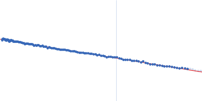
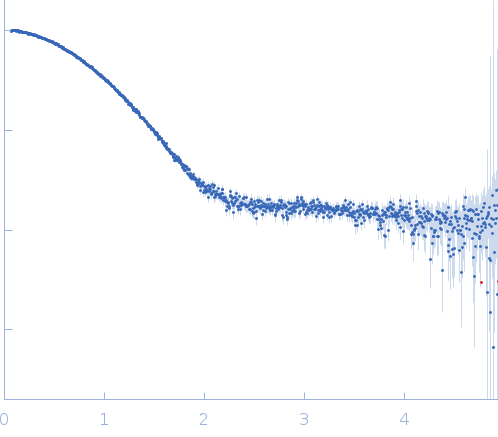
Synchrotron SAXS data from solutions of S2MH1E3 in 20 mM Tris 150 mM NaCl, pH 7.2 were collected on the BM29 beam line at the ESRF (Grenoble, France) using a Pilatus 1M detector at a wavelength of λ = 0.099 nm (I(s) vs s, where s = 4πsinθ/λ, and 2θ is the scattering angle). Solute concentrations ranging between 1 and 5 mg/ml were measured at 10°C. The data were normalized to the intensity of the transmitted beam and radially averaged; the scattering of the solvent-blank was subtracted. The low angle data collected at lower concentration were merged with the highest concentration high angle data to yield the final composite scattering curve.
Sample detector distance = UNKNOWN. X-ray Exposure time = UNKNOWN. Number of frames = UNKNOWN
 s, nm-1