Molecular basis for metabolite channeling in a ring opening enzyme of the phenylacetate degradation pathway.

Sathyanarayanan N, Cannone G, Gakhar L, Katagihallimath N, Sowdhamini R, Ramaswamy S, Vinothkumar KR, Nat Commun 10(1):4127 (2019) Europe PMC

SASDGL2 – Ring opening PaaZ from the phenylacetate degradation pathway (E. coli K12)

Bifunctional protein PaaZ
MWexperimental 434 kDa
MWexpected 438 kDa
VPorod 636 nm3
log I(s) 6.48×10³ 6.48×10² 6.48×101 6.48×100
Bifunctional protein PaaZ small angle scattering data  s, nm-1
ln I(s)
Bifunctional protein PaaZ Guinier plot ln 6.48×10³ Rg: 6.2 nm 0 (6.2 nm)-2 s2
(sRg)2I(s)/I(0)
Bifunctional protein PaaZ Kratky plot 1.104 0 3 sRg
p(r)
Bifunctional protein PaaZ pair distance distribution function Rg: 6.2 nm 0 Dmax: 20.0 nm

Data validation


Fits and models


log I(s)
 s, nm-1
Bifunctional protein PaaZ OTHER model

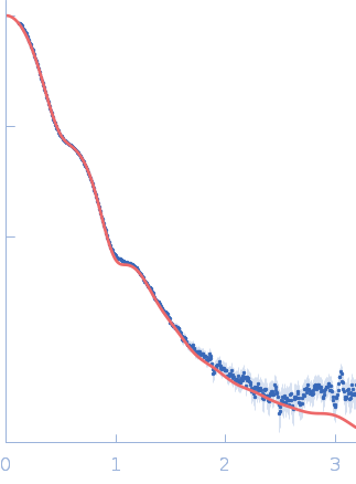
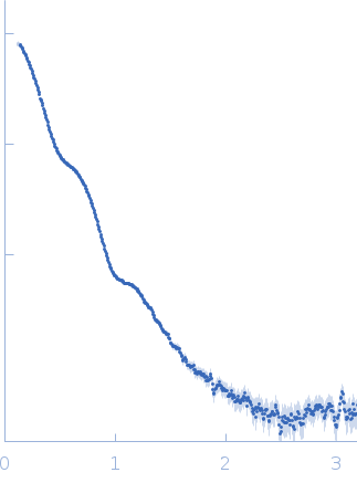
Synchrotron SAXS data from solutions of the bifunctional protein PaaZ in 25 mM HEPES, 50 mM NaCl, pH 7.4 were collected on the 12.3.1 (SIBYLS) beam line at the Advanced Light Source (ALS; Berkeley, CA, USA) using a MAR 165 CCD detector at a sample-detector distance of 1.5 m and at a wavelength of λ = 0.103 nm (I(s) vs s, where s = 4πsinθ/λ, and 2θ is the scattering angle). Solute concentrations ranging between 1.5 and 4.5 mg/ml were measured at 10°C. One 5 second frame was collected per concentration. The data were normalized to the intensity of the transmitted beam and radially averaged; the scattering of the solvent-blank was subtracted. The low angle data collected at lower concentration were merged with the highest concentration high angle data to yield the final composite scattering curve.

Bifunctional protein PaaZ (PaaZ)
Mol. type   Protein
Organism   Escherichia coli
Olig. state   Hexamer
Mon. MW   73.0 kDa
 
UniProt   P77455 (1-681)
Sequence   FASTA
 
PDB ID   6JQL