Combination of SAXS and Protein Painting Discloses the Three-Dimensional Organization of the Bacterial Cysteine Synthase Complex, a Potential Target for Enhancers of Antibiotic Action.

Rosa B, Marchetti M, Paredi G, Amenitsch H Franko N, Benoni R, Giabbai B, De Marino MG, Mozzarelli A, Ronda L, Storici P, Campanini B, Bettati S, Int J Mol Sci 20(20) (2019) Europe PMC

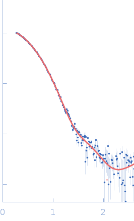
SASDGV6 – Cysteine synthase A

Cysteine synthase A
MWexperimental 72 kDa
MWexpected 71 kDa
VPorod 108 nm3
log I(s) 6.71×100 6.71×10⁻¹ 6.71×10⁻² 6.71×10⁻³
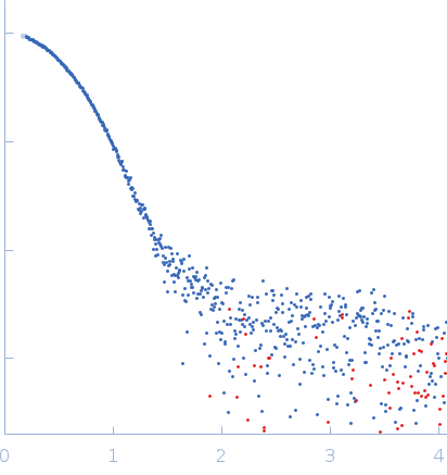
Cysteine synthase A small angle scattering data  s, nm-1
ln I(s)
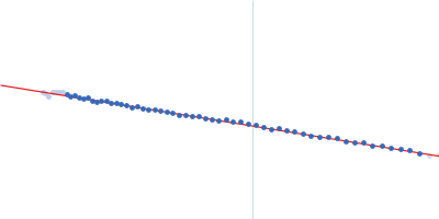
Cysteine synthase A Guinier plot ln 6.71×100 Rg: 2.6 nm 0 (2.6 nm)-2 s2
(sRg)2I(s)/I(0)
Cysteine synthase A Kratky plot 1.104 0 3 sRg
p(r)
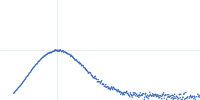
Cysteine synthase A pair distance distribution function Rg: 2.6 nm 0 Dmax: 8.5 nm

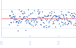
Data validation


Fits and models


log I(s)
 s, nm-1
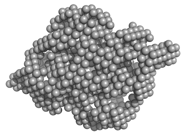
Cysteine synthase A DAMMIN model

Synchrotron SAXS data from solutions of cysteine synthase in 20 mM sodium phosphate, 85 mM NaCl, 2 mM EDTA, 10 mM 2-MCE, pH 7.5 were collected on the Austrian SAXS beamline 5.2L beam line at the ELETTRA Sincrotrone Trieste storage ring (Trieste, Italy) using a Dectris / Pilatus3 1M detector at a sample-detector distance of 1.2 m and at a wavelength of λ = 0.15 nm (I(s) vs s, where s = 4πsinθ/λ, and 2θ is the scattering angle). One solute concentration of 1.20 mg/ml was measured at 20°C. 12 successive 10 second frames were collected. The data were normalized to the intensity of the transmitted beam and radially averaged; the scattering of the solvent-blank was subtracted.

Cysteine synthase A (CysK)
Mol. type   Protein
Organism   Escherichia coli
Olig. state   Dimer
Mon. MW   35.7 kDa
 
UniProt   P0ABK5 (1-323)
Sequence   FASTA