|
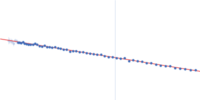
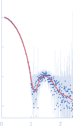
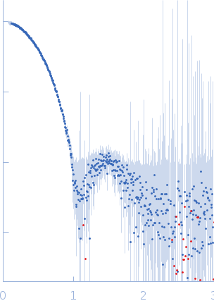
Synchrotron SAXS data from solutions of Uricase in 20 mM Tris. 150 mM NaCl, 1 mM EDTA, 5 mM DTT, pH 8, were collected on the BL4-2 beam line at the Stanford Synchrotron Radiation Lightsource (SSRL; Menlo Park, CA, USA) using a Pilatus3 X 1M detector at a sample-detector distance of 1.7 m and at a wavelength of λ = 0.112709 nm (I(s) vs s, where s = 4πsinθ/λ, and 2θ is the scattering angle). In-line size-exclusion chromatography (SEC) SAS was employed. The SEC parameters were as follows: A 50 μl sample at 5 mg/ml was injected at a 0.05 ml/min flow rate onto a GE Superdex 200 Increase 3.2/300 column at 23°C. 500 successive 1 second frames were collected throughout the entire SEC run. The data were normalized to the intensity of the transmitted beam and radially averaged; the scattering of an appropriate solvent-blank (from the SEC column eluate) were subtracted from the appropriate sample peak frames.
|
|
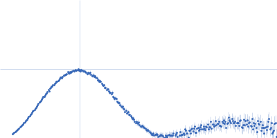
 s, nm-1
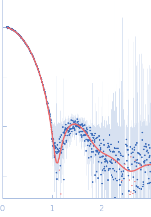