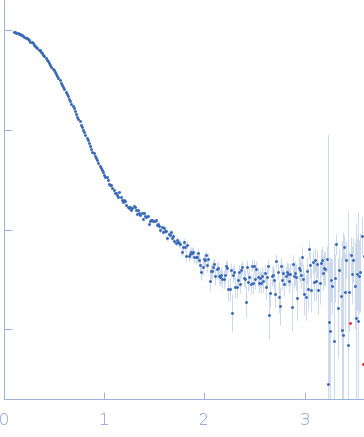
Synchrotron SAXS data from solutions of Apolipoprotein-D oxidised with H2O2 in 50 mM Na Phosphate, 150 mM NaCl, 3% glycerol, pH 7.4 were collected on the SAXS/WAXS beam line at the Australian Synchrotron (Melbourne, Australia) using a Pilatus 1M detector at a wavelength of λ = 0.10322 nm (I(s) vs s, where s = 4πsinθ/λ, and 2θ is the scattering angle). In-line size-exclusion chromatography co-flow (SEC) SAS was employed. The SEC parameters were as follows: A 100.00 μl sample at 9.4 mg/ml was injected at a 0.45 ml/min flow rate onto a GE Superdex 200 Increase 5/150 column . 492 successive 1 second frames were collected. The data were normalized to the intensity of the transmitted beam and radially averaged; the scattering of the solvent-blank was subtracted.
Apolipoprotein-D (43.45 µM) was incubated with H2O2 (final concentration 100 mM overnight at 4°C. The sample was spin-concentrated using an Amicon Ultra concentrator with an Ultracel-10 membrane and frozen at −80°C. 100 µl of concentrated sample (9.40 mg/ml) was applied to the SEC column.
 s, nm-1