| MWexperimental | 20 | kDa |
| MWexpected | 20 | kDa |
| VPorod | 33 | nm3 |
|
log I(s)
2.53×100
2.53×10⁻¹
2.53×10⁻²
2.53×10⁻³
|
 s, nm-1
|
|
|
|

|
|

|
|

|
|
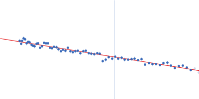
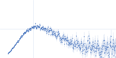
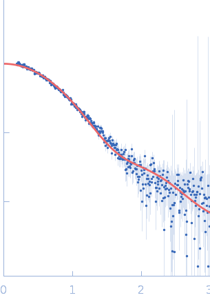
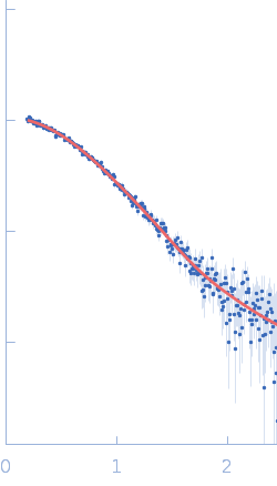
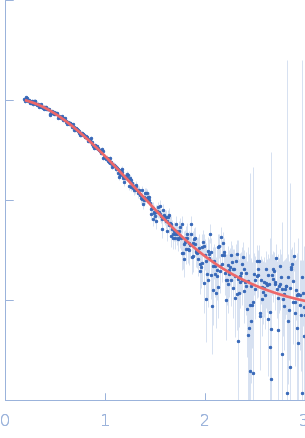
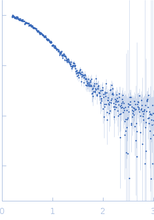
SAXS data from solutions of Protein ninH (T15A) in 150 mM NaCl, 50 mM phosphate buffer, pH 7.4, 1 mM EDTA were collected using Xenocs Xeuss 2.0 instrument (Department of Macromolecular Physics, Adam Mickiewicz University, Poznań, Poland) equipped with a MetalJet D2 microfocus X-ray generator and a Pilatus 3R 1M detector at a wavelength of λ = 0.134 nm (I(s) vs s, where s = 4πsinθ/λ, and 2θ is the scattering angle). A sample was collected from a SEC column immediately prior to batch analysis. One solute concentration of 4.00 mg/ml was measured at 22°C. Three successive 600 second frames were collected. The data were normalized to the intensity of the transmitted beam and radially averaged and each exposure checked for radiation damage; the scattering of the solvent-blank was subtracted.
The resulting SAXS data used to validate the model of NinH derived from MODELLER. |
|
|||||||||||||||||||||||||||