Structure, Dynamics and Cellular Insight Into Novel Substrates of the Legionella pneumophila Type II Secretion System

Portlock T, Tyson J, Dantu S, Rehman S, White R, McIntire I, Sewell L, Richardson K, Shaw R, Pandini A, Cianciotto N, Garnett J, Frontiers in Molecular Biosciences 7 (2020) DOI

SASDHW2 – The Legionella pneumophila type II secretion system substrate NttE

Uncharacterized protein
MWexperimental 66 kDa
MWexpected 66 kDa
VPorod 114 nm3
log I(s) 3.79×10⁻² 3.79×10⁻³ 3.79×10⁻⁴ 3.79×10⁻⁵
Uncharacterized protein small angle scattering data  s, nm-1
ln I(s)
Uncharacterized protein Guinier plot ln 3.79×10⁻² Rg: 2.9 nm 0 (2.9 nm)-2 s2
(sRg)2I(s)/I(0)
Uncharacterized protein Kratky plot 1.104 0 3 sRg
p(r)
Uncharacterized protein pair distance distribution function Rg: 2.9 nm 0 Dmax: 9.4 nm

Data validation


Fits and models


log I(s)
 s, nm-1
Uncharacterized protein GASBOR model

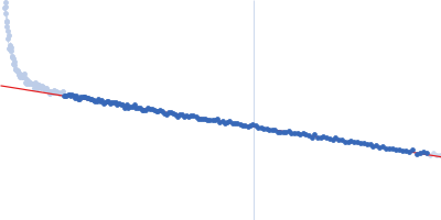
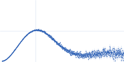
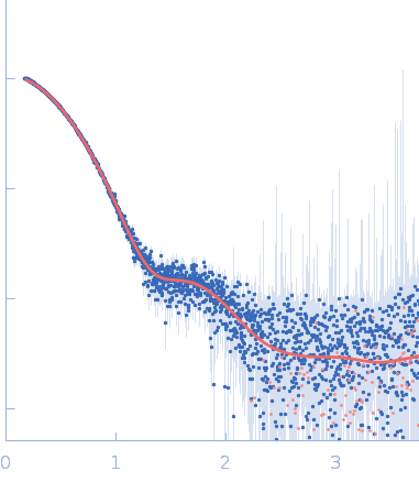
Synchrotron SAXS data from solutions of the Legionella pneumophila type II secretion system substrate NttE in 20 mM Tris 200 mM NaCl, pH 8 were collected on the B21 beam line at the Diamond Light Source (Didcot, UK) using a Pilatus 2M detector at a wavelength of λ = 0.1 nm (I(s) vs s, where s = 4πsinθ/λ, and 2θ is the scattering angle). In-line size-exclusion chromatography (SEC) SAS was employed. The SEC parameters were as follows: A 60.00 μl sample at 10 mg/ml was injected at a 0.16 ml/min flow rate onto a Shodex KW403 column at 25°C. The data were normalized to the intensity of the transmitted beam and radially averaged; the scattering of an appropriate the solvent-blank was subtracted from the scattering data obtained through the sample SEC-elution peak.

Sample detector distance = UNKNOWN. X-ray Exposure time = UNKNOWN. Number of frames = UNKNOWN

Uncharacterized protein
Mol. type   Protein
Organism   Legionella pneumophila
Olig. state   Dimer
Mon. MW   33.2 kDa
 
UniProt   A0A4Q5N6R9 (20-288)
Sequence   FASTA