An atypical BRCT-BRCT interaction with the XRCC1 scaffold protein compacts human DNA Ligase IIIα within a flexible DNA repair complex.

Hammel M Rashid I, Sverzhinsky A, Pourfarjam Y, Tsai MS, Ellenberger T, Pascal JM, Kim IK, Tainer JA, Tomkinson AE, Nucleic Acids Res (2020) Europe PMC

SASDJ72 – DNA Ligase IIIα monomer/dimer

DNA ligase 3 (DNA ligase III alpha)
MWexperimental 150 kDa
VPorod 240 nm3
log I(s) 1.69×10² 1.69×101 1.69×100 1.69×10⁻¹
DNA ligase 3 (DNA ligase III alpha) small angle scattering data  s, nm-1
ln I(s)
DNA ligase 3 (DNA ligase III alpha) Guinier plot ln 1.69×10² Rg: 6.1 nm 0 (6.1 nm)-2 s2
(sRg)2I(s)/I(0)
DNA ligase 3 (DNA ligase III alpha) Kratky plot 1.104 0 3 sRg
p(r)
DNA ligase 3 (DNA ligase III alpha) pair distance distribution function Rg: 6.3 nm 0 Dmax: 21 nm

Data validation


Fits and models


log I(s)
 s, nm-1
DNA ligase 3 (DNA ligase III alpha) BILBOMD model

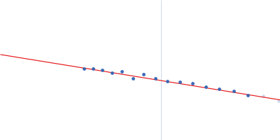
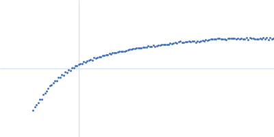
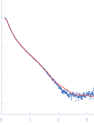
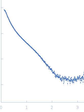
Synchrotron SAXS data from solutions of DNA Ligase IIIα in 25 mM Tris-HCl pH 7.5, 150 mM NaCl, 10% glycerol, 2 mM DTT, were collected on the 12.3.1 (SIBYLS) beam line at the Advanced Light Source (ALS; Berkeley, CA, USA) using a MAR 165 CCD detector at a sample-detector distance of 1.5 m and at a wavelength of λ = 0.11 nm (I(s) vs s, where s = 4πsinθ/λ, and 2θ is the scattering angle). Solute concentrations ranging between 1 and 5 mg/ml were measured at 20°C. Three successive 0.500 second frames were collected. The data were normalized to the intensity of the transmitted beam and radially averaged; the scattering of the solvent-blank was subtracted. The low angle data collected at lower concentration were merged with the highest concentration high angle data to yield the final composite scattering curve.

DNA ligase 3 (DNA ligase III alpha) (LigIIIα)
Mol. type   Protein
Organism   Homo sapiens
Olig. state   Other
Mon. MW   102.7 kDa
 
UniProt   P49916 (88-1009)
Sequence   FASTA