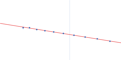
| MWI(0) | 368 | kDa |
| MWexpected | 446 | kDa |
|
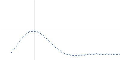
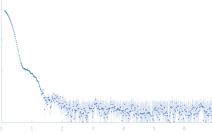
log I(s)
3.66×10⁵
3.66×10⁴
3.66×10³
3.66×10²
|
 s, nm-1
|
|
|
|

|
|


|
|
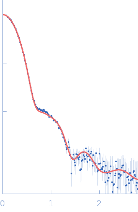
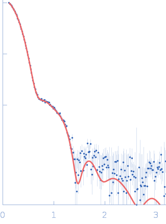
SAXS
data from solutions of
Lactococcus phage Phi28 apo-gp11 encapsidation protein
in
100 mM NaCl, 25 mM HEPES, pH 7.4
were collected
using a Pilatus 100K detector
at a wavelength of λ = 1.5418 nm
(I(s) vs s, where s = 4πsinθ/λ, and 2θ is the scattering angle).
One solute concentration of 1.00 mg/ml was measured
at 10°C.
12 successive
3600 second frames were collected.
The data were normalized to the intensity of the transmitted beam and radially averaged; the scattering of the solvent-blank was subtracted.
Sample detector distance = UNKNOWN |
|
|||||||||||||||||||||||||||||||||