Inference of molecular structure for characterization and improvement of clinical grade immunocytokines

Ongaro T, Guarino S, Scietti L, Palamini M, Wulhfard S, Neri D, Villa A, Forneris F, Journal of Structural Biology :107696 (2021) DOI

SASDJN8 – L19-IL2 dimeric immunocytokine

L19-IL2 dimeric immunocytokine
MWI(0) 64 kDa
MWexpected 84 kDa
VPorod 128 nm3
log I(s) 6.62×101 6.62×100 6.62×10⁻¹ 6.62×10⁻²
L19-IL2 dimeric immunocytokine small angle scattering data  s, nm-1
ln I(s)
L19-IL2 dimeric immunocytokine Guinier plot ln 6.62×101 Rg: 4.1 nm 0 (4.1 nm)-2 s2
(sRg)2I(s)/I(0)
L19-IL2 dimeric immunocytokine Kratky plot 1.104 0 3 sRg
p(r)
L19-IL2 dimeric immunocytokine pair distance distribution function Rg: 4.1 nm 0 Dmax: 11.9 nm

Data validation


Fits and models


log I(s)
 s, nm-1
L19-IL2 dimeric immunocytokine ALLOSMOD model

log I(s)
 s, nm-1
L19-IL2 dimeric immunocytokine GASBOR model

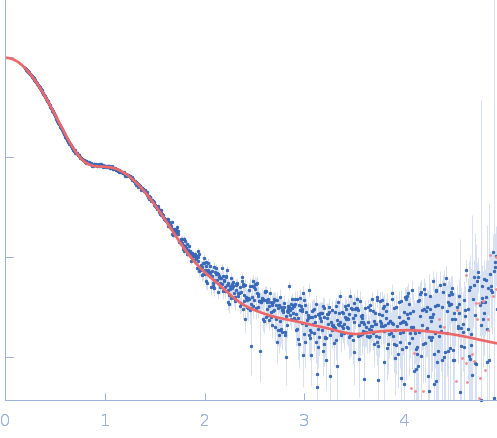
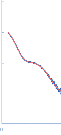
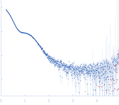
Synchrotron SAXS data from solutions of the L19-IL2 immunocytokine in 25 mM HEPES/NaOH, 0.2 M NaCl, pH 8 were collected on the BM29 beam line at the ESRF (Grenoble, France) using a Pilatus 1M detector at a sample-detector distance of 2.9 m and at a wavelength of λ = 0.099 nm (I(s) vs s, where s = 4πsinθ/λ, and 2θ is the scattering angle). Size exclusion chromatography coupled to SAXS (SEC-SAXS) was carried out at at 20 °C using Nexera High Pressure Liquid Chromatography system (HPLC; Shimadzu). 50 μL of L19-IL2 concentrated at 4 mg/mL were injected into a Superdex 200 3.2/300 PC (GE Healthcare), pre-equilibrated with 25 mM HEPES/NaOH, 200 mM NaCl, pH 8.0. 101 successive 1 second frames were collected through the sample elution peak and processed using CHROMIXS that included the identification and subtraction of an appropriate solvent-blank.

L19-IL2 dimeric immunocytokine (L19-IL2)
Mol. type   Protein
Organism   synthetic construct
Olig. state   Dimer
Mon. MW   42.0 kDa
Sequence   FASTA