| MWI(0) | 43 | kDa |
| MWexpected | 47 | kDa |
| VPorod | 28 | nm3 |
|
log I(s)
1.07×10²
1.07×101
1.07×100
1.07×10⁻¹
|
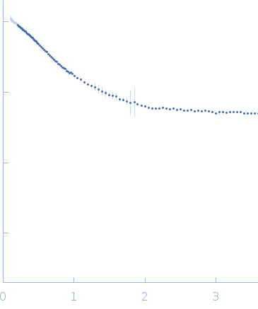 s, nm-1
|
|
|
|

|
|

|
|

|
|
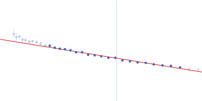
Synchrotron SAXS
data from solutions of
Interferon-activable protein 204 from Mus musculus (Mouse) amino-acids 215-619
in
20 mM HEPES, 100 mM KCl, pH 7.4
were collected
on the
X9A beam line
at the National Synchrotron Light Source (NSLS) storage ring
(Brookhaven, NY, USA)
using a Pilatus 300K detector
at a wavelength of λ = 0.1033 nm
(I(s) vs s, where s = 4πsinθ/λ, and 2θ is the scattering angle).
One solute concentration of 3.80 mg/ml was measured
at 25°C.
30 successive
2.300 second frames were collected.
The data were normalized to the intensity of the transmitted beam and radially averaged; the scattering of the solvent-blank was subtracted.
The low angle data collected at lower concentration were merged with the highest concentration high angle data to yield the final composite scattering curve.
Sample detector distance = UNKNOWN. Concentration min = UNKNOWN |
|
|||||||||||||||||||||||||||