Mechanistic insights into RNA surveillance by the canonical poly(A) polymerase Pla1 of the MTREC complex.

Soni K Sivadas A, Horvath A, Dobrev N, Hayashi R, Kiss L, Simon B, Wild K, Sinning I, Fischer T, Nat Commun 14(1):772 (2023) Europe PMC

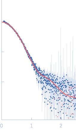
SASDKE6 – Poly(A) polymerase pla1 bound to the NURS complex subunit red1

NURS complex subunit red1
Poly(A) polymerase pla1
MWexperimental 86 kDa
MWexpected 73 kDa
VPorod 138 nm3
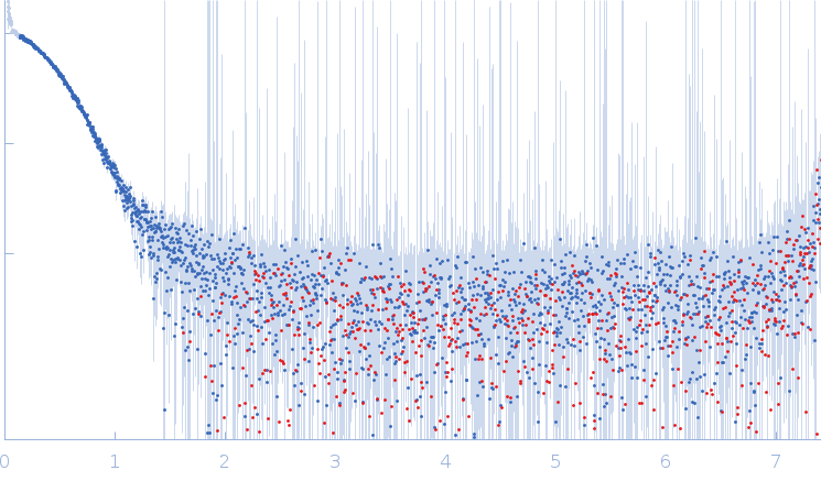
log I(s) 1.92×10⁴ 1.92×10³ 1.92×10² 1.92×101
NURS complex subunit red1 Poly(A) polymerase pla1 small angle scattering data  s, nm-1
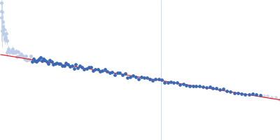
ln I(s)
NURS complex subunit red1 Poly(A) polymerase pla1 Guinier plot ln 1.93×10⁴ Rg: 3.3 nm 0 (3.3 nm)-2 s2
(sRg)2I(s)/I(0)
NURS complex subunit red1 Poly(A) polymerase pla1 Kratky plot 1.104 0 3 sRg
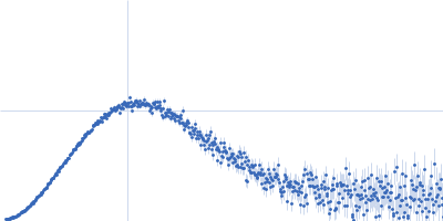
p(r)
NURS complex subunit red1 Poly(A) polymerase pla1 pair distance distribution function Rg: 3.3 nm 0 Dmax: 10.7 nm

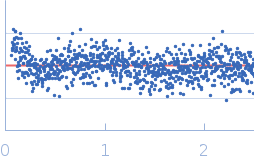
Data validation


Fits and models


log I(s)
 s, nm-1
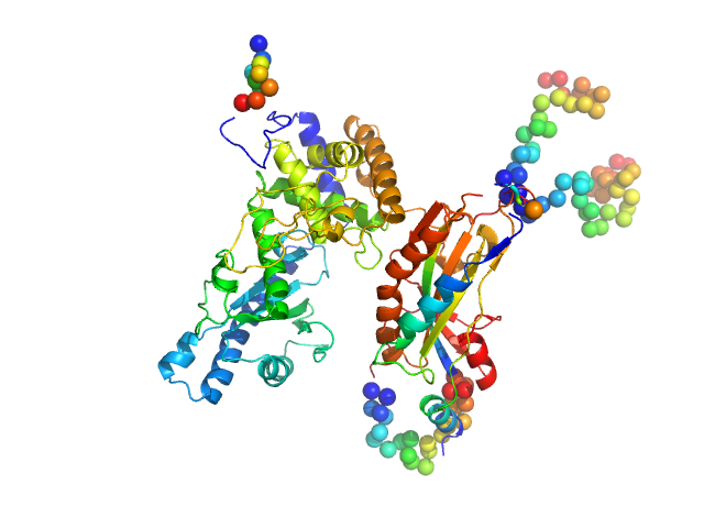
NURS complex subunit red1 Poly(A) polymerase pla1 CORAL model

Synchrotron SAXS data from solutions the pla1-red1 complex in 20 mM HEPES, pH 7.5, 150 mM NaCl, 1mM DTT, were collected on the EMBL P12 beam line at PETRA III (DESY, Hamburg, Germany) using a Pilatus 6M detector at a sample-detector distance of 3 m and at a wavelength of λ = 0.124193 nm (I(s) vs s, where s = 4πsinθ/λ, and 2θ is the scattering angle). One solute concentration of 0.85 mg/ml was measured at 20°C. 40 successive 0.195 second frames were collected. The data were normalized to the intensity of the transmitted beam and radially averaged; the scattering of the solvent-blank was subtracted.

NURS complex subunit red1 (Red1)
Mol. type   Protein
Organism   Schizosaccharomyces pombe (strain 972 / ATCC 24843)
Olig. state   Monomer
Mon. MW   7.6 kDa
 
UniProt   Q9UTR8 (288-345)
Sequence   FASTA
 
Poly(A) polymerase pla1 (Pla1)
Mol. type   Protein
Organism   Schizosaccharomyces pombe (strain 972 / ATCC 24843)
Olig. state   Monomer
Mon. MW   65.3 kDa
 
UniProt   Q10295 (1-566)
Sequence   FASTA