| MWexperimental | 79 | kDa |
| MWexpected | 77 | kDa |
| VPorod | 110 | nm3 |
|
log I(s)
9.30×100
9.30×10⁻¹
9.30×10⁻²
9.30×10⁻³
|
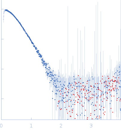 s, nm-1
|
|
|
|

|
|

|
|

|
|
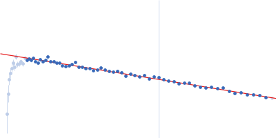
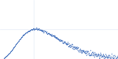
SAXS data from solutions of Malus domestica double bond reductase (MdDBR), apoform, in 50 mM Tris-HCl, 100 mM NaCl., pH 7.5 were collected using an Anton Paar SAXSpoint 2.0 instrument equipped with a Eiger R 1M detector (Vestec, Czech Republic) at a sample-detector distance of 0.8 m and at a wavelength of λ = 0.134 nm (I(s) vs s, where s = 4πsinθ/λ, and 2θ is the scattering angle). One solute concentration of 7.00 mg/ml was measured at 20°C. 30 successive 60 second frames were collected. The data were normalized to the intensity of the transmitted beam and radially averaged; the scattering of the solvent-blank was subtracted.
|
|
|||||||||||||||||||||||||||||||||