The structural and functional characterization of Malus domestica double bond reductase MdDBR provides insights towards the identification of its substrates

Caliandro R, Polsinelli I Demitri N, Musiani F, Martens S, Benini S, International Journal of Biological Macromolecules 171:89-99 (2021) DOI

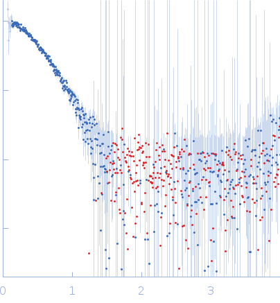
SASDKH4 – Malus domestica double bond reductase in the presence of NADPH 5mM

Malus domestica double bond reductase
MWexperimental 91 kDa
MWexpected 77 kDa
VPorod 99 nm3
log I(s) 2.65×100 2.65×10⁻¹ 2.65×10⁻² 2.65×10⁻³
Malus domestica double bond reductase small angle scattering data  s, nm-1
ln I(s)
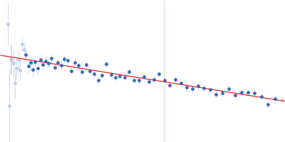
Malus domestica double bond reductase Guinier plot ln 2.65×100 Rg: 3.1 nm 0 (3.1 nm)-2 s2
(sRg)2I(s)/I(0)
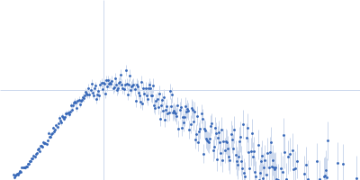
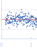
Malus domestica double bond reductase Kratky plot 1.104 0 3 sRg
p(r)
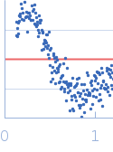
Malus domestica double bond reductase pair distance distribution function Rg: 3.2 nm 0 Dmax: 10.7 nm

Data validation


Fits and models


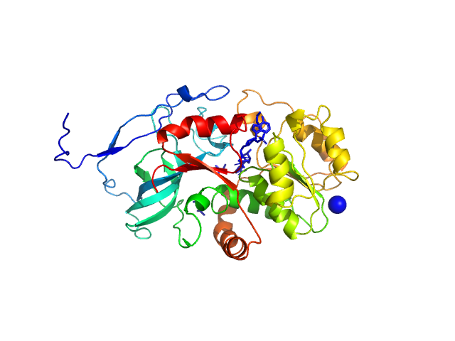
log I(s)
 s, nm-1
Malus domestica double bond reductase PYMOL model

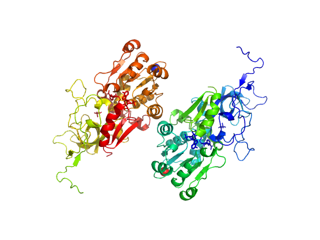
log I(s)
 s, nm-1
Malus domestica double bond reductase PYMOL model

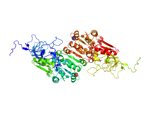
log I(s)
 s, nm-1
Malus domestica double bond reductase PYMOL model

SAXS data from solutions of Malus domestica double bond reductase in 50 mM Tris-HCl, 100 mM NaCl, 5 mM NADPH, pH 7.5 were collected using an Anton Paar SAXSpoint 2.0 instrument equipped with a Eiger R 1M detector (Vestec, Czech Republic) at a sample-detector distance of 0.8 m and at a wavelength of λ = 0.134 nm (I(s) vs s, where s = 4πsinθ/λ, and 2θ is the scattering angle). One solute concentration of 2.20 mg/ml was measured at 20°C. 30 successive 60 second frames were collected. The data were normalized to the intensity of the transmitted beam and radially averaged; the scattering of the solvent-blank was subtracted.

Malus domestica double bond reductase (MdDBR)
Mol. type   Protein
Organism   Malus domestica
Olig. state   Dimer
Mon. MW   38.4 kDa
Sequence   FASTA
 
PDB ID   6YTZ