Liang W,
Wijaya J,
Wei H,
Noble A,
Mancl J,
Mo S,
Lee D,
Lin King J,
Pan M,
Liu C,
Koehler C,
Zhao M,
Potter C,
Carragher B,
Li S,
Tang W,
Nature Communications
13(1)
(2022)
DOI
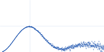
Synchrotron SAXS
data from solutions of
SEC-SAXS of Presequence Protease (PreP)
in
20 mM Tris, 100 mM NaCl, pH 7.7
were collected
on the
BioCAT 18ID beam line
at the Advanced Photon Source (APS), Argonne National Laboratory storage ring
(Lemont, IL, USA)
using a Pilatus 100K detector
at a sample-detector distance of 3.7 m and
at a wavelength of λ = 0.10332 nm
(I(s) vs s, where s = 4πsinθ/λ, and 2θ is the scattering angle).
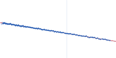
In-line size-exclusion chromatography (SEC) SAS was employed. The SEC parameters were as follows: A sample
was injected at a 1.40 ml/min flow rate
onto a GE Superdex 200 10/300 column
at 22°C.
One
0.500 second frame was collected.
The data were normalized to the intensity of the transmitted beam and radially averaged; the scattering of the solvent-blank was subtracted.
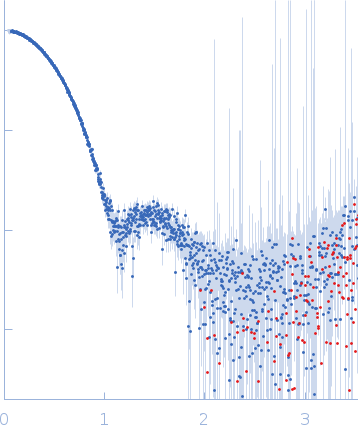 s, nm-1