An integrative NMR-SAXS approach for structural determination of large RNAs defines the substrate-free state of a trans -cleaving Neurospora Varkud Satellite ribozyme

Dagenais P, Desjardins G, Legault P, Nucleic Acids Research (2021) DOI

SASDKV3 – III-IV-V three-way junction from the Neurospora Varkud Satellite (VS) ribozyme

Neurospora Varkud Satellite ribozyme junction III-IV-V
MWexperimental 18 kDa
MWexpected 15 kDa
VPorod 20 nm3
log I(s) 7.39×100 7.39×10⁻¹ 7.39×10⁻² 7.39×10⁻³
Neurospora Varkud Satellite ribozyme junction III-IV-V small angle scattering data  s, nm-1
ln I(s)
Neurospora Varkud Satellite ribozyme junction III-IV-V Guinier plot ln 7.39×100 Rg: 2.1 nm 0 (2.1 nm)-2 s2
(sRg)2I(s)/I(0)
Neurospora Varkud Satellite ribozyme junction III-IV-V Kratky plot 1.104 0 3 sRg
p(r)
Neurospora Varkud Satellite ribozyme junction III-IV-V pair distance distribution function Rg: 2.1 nm 0 Dmax: 7.5 nm

Data validation


Fits and models


log I(s)
 s, nm-1
Neurospora Varkud Satellite ribozyme junction III-IV-V OTHER model

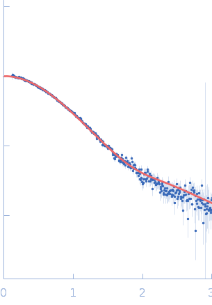
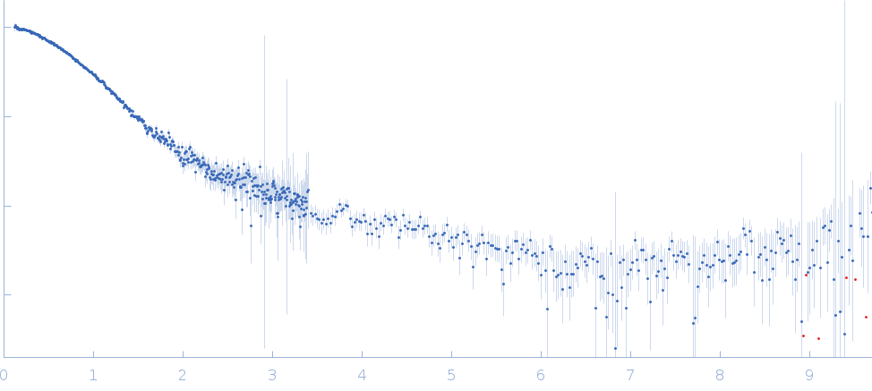
Synchrotron SAXS data from solutions of III-IV-V three-way junction from the VS ribozyme in 50 mM MES, 50 mM KCl, 5 mM MgCl2, pH 6.5 were collected on the G1 beam line at the Cornell High Energy Synchrotron Source (CHESS; Ithaca, NY, USA) using a Pilatus 100K detector at a sample-detector distance of 1.5 m and at a wavelength of λ = 0.1245 nm (I(s) vs s, where s = 4πsinθ/λ, and 2θ is the scattering angle). Solute concentrations ranging between 0.5 and 1 mg/ml were measured at 20°C. 10 successive 2 second frames were collected. The data were normalized to the intensity of the transmitted beam and radially averaged; the scattering of the solvent-blank was subtracted. The low angle data collected at lower concentration were merged with the highest concentration high angle data to yield the final composite scattering curve.

Neurospora Varkud Satellite ribozyme junction III-IV-V (VS ribozyme J345)
Mol. type   RNA
Organism   Neurospora crassa
Olig. state   Monomer
Mon. MW   15.2 kDa
Sequence   FASTA