Despite the odds: formation of the SARS-CoV-2 methylation complex

Matsuda A, Plewka J, Rawski M, Mourão A, Zajko W, Siebenmorgen T, Kresik L, Lis K, Jones A, Pachota M, Karim A, Hartman K, Nirwal S, Sonani R, Chykunova Y, Minia I, Mak P, Landthaler M, Nowotny M, Dubin G, Sattler M, Suder P, Popowicz G, Pyrć K, Czarna A, Nucleic Acids Research (2024) DOI

SASDKW6 – SARS-CoV-2 non-structural protein 10/non-structural protein 14/non-structural protein 16 triplex (nsp10/nsp14/nsp16)

Replicase polyprotein 1ab (non-structural protein 14)
Replicase polyprotein 1a (non-structural protein 10)
Replicase polyprotein 1ab (non-structural protein 16)
MWexperimental 86 kDa
MWexpected 108 kDa
VPorod 113 nm3
log I(s) 5.83×100 5.83×10⁻¹ 5.83×10⁻² 5.83×10⁻³
Replicase polyprotein 1ab (non-structural protein 14) Replicase polyprotein 1a (non-structural protein 10) Replicase polyprotein 1ab (non-structural protein 16) small angle scattering data  s, nm-1
ln I(s)
Replicase polyprotein 1ab (non-structural protein 14) Replicase polyprotein 1a (non-structural protein 10) Replicase polyprotein 1ab (non-structural protein 16) Guinier plot ln 5.83×100 Rg: 4.7 nm 0 (4.7 nm)-2 s2
(sRg)2I(s)/I(0)
Replicase polyprotein 1ab (non-structural protein 14) Replicase polyprotein 1a (non-structural protein 10) Replicase polyprotein 1ab (non-structural protein 16) Kratky plot 1.104 0 3 sRg
p(r)
Replicase polyprotein 1ab (non-structural protein 14) Replicase polyprotein 1a (non-structural protein 10) Replicase polyprotein 1ab (non-structural protein 16) pair distance distribution function Rg: 3.7 nm 0 Dmax: 11.4 nm

Data validation


Fits and models


log I(s)
 s, nm-1
Replicase polyprotein 1ab (non-structural protein 14) Replicase polyprotein 1a (non-structural protein 10) Replicase polyprotein 1ab (non-structural protein 16) GASBOR model

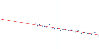
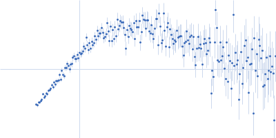
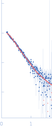
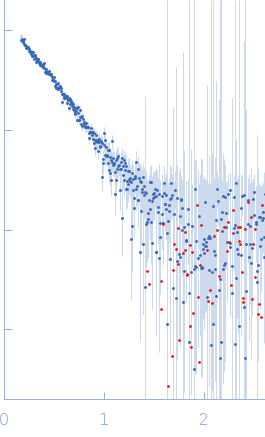
Synchrotron SAXS data from solutions of the SARS-CoV-2 nsp10/nsp14/nsp16 complex in 50 mM Tris, 150 mM NaCl, 5 mM MgCl2, 2 mM β-mercaptoethanol, pH 8.5 were collected on the BM29 beam line at the ESRF (Grenoble, France) using a Pilatus 1M detector at a sample-detector distance of 2.8 m and at a wavelength of λ = 0.1 nm (I(s) vs s, where s = 4πsinθ/λ, and 2θ is the scattering angle). In-line size-exclusion chromatography (SEC) SAS was employed. The SEC parameters were as follows: A 100.00 μl sample at 2.6 mg/ml was injected onto a Agilent Bio SEC-3, 300 Å column. The data were normalized to the intensity of the transmitted beam and radially averaged; the scattering of the solvent-blank was subtracted.

CAUTION: A significant discrepancy is noted between the experimentally determined (86 kDa) vs. expected (108 kDa) molecular weights. CAUTION! The Porod volume (113 nm^3) is inconsistent with the formation of a 108 kDa complex (expected Porod volume = 173 nm^3). Experimental temperature: UNKNOWN. X-ray exposure time: UNKNOWN. Number of data frames collected: UNKNOWN. SEC-flow rate: UNKNOWN.

Replicase polyprotein 1ab (non-structural protein 14) (nsp14)
Mol. type   Protein
Organism   Severe acute respiratory syndrome coronavirus 2
Olig. state   Monomer
Mon. MW   60.1 kDa
 
UniProt   P0DTD1 (5926-6452)
Sequence   FASTA
 
Replicase polyprotein 1a (non-structural protein 10) (nsp10)
Mol. type   Protein
Organism   Severe acute respiratory syndrome coronavirus 2
Olig. state   Monomer
Mon. MW   15.0 kDa
 
UniProt   P0DTC1 (4254-4392)
Sequence   FASTA
 
Replicase polyprotein 1ab (non-structural protein 16) (nsp16)
Mol. type   Protein
Organism   Severe acute respiratory syndrome coronavirus 2
Olig. state   Monomer
Mon. MW   33.4 kDa
 
UniProt   P0DTD1 (6799-7096)
Sequence   FASTA