A fragment-based approach identifies an allosteric pocket that impacts malate dehydrogenase activity

Reyes Romero A, Lunev S, Popowicz G, Calderone V, Gentili M, Sattler M, Plewka J Taube M, Kozak M, Holak T, Dömling A, Groves M, Communications Biology 4(1) (2021) DOI

SASDLS2 – PfMDH L-lactate dehydrogenase bound to inhibitor 6a

L-lactate dehydrogenase
MWexperimental 114 kDa
MWexpected 141 kDa
VPorod 223 nm3
log I(s) 4.46×100 4.46×10⁻¹ 4.46×10⁻² 4.46×10⁻³
L-lactate dehydrogenase small angle scattering data  s, nm-1
ln I(s)
L-lactate dehydrogenase Guinier plot ln 4.47×100 Rg: 3.6 nm 0 (3.6 nm)-2 s2
(sRg)2I(s)/I(0)
L-lactate dehydrogenase Kratky plot 1.104 0 3 sRg
p(r)
L-lactate dehydrogenase pair distance distribution function Rg: 3.5 nm 0 Dmax: 11.4 nm

Data validation


Fits and models


log I(s)
 s, nm-1
L-lactate dehydrogenase DAMMIF model

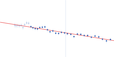
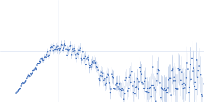
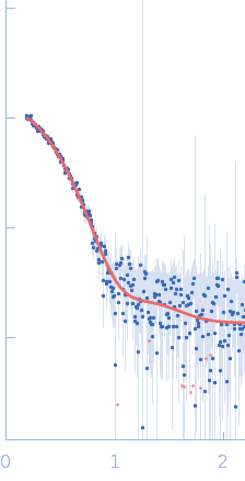
SAXS data from solutions of PfMDH L-lactate dehydrogenase bound to inhibitor 6a in 100 mM Na-phosphate buffer, 400 mM NaCl, pH 7.4 were measured using a Xenocs Xeuss 2.0 instrument equipped with a Pilatus3 R 1M detector (Department of Macromolecular Physics, Adam Mickiewicz University, Poznań, Poland) at a wavelength of λ = 0.134 nm (I(s) vs s, where s = 4πsinθ/λ, and 2θ is the scattering angle). One solute concentration of 2.49 mg/ml was measured. Two successive 600 second frames were collected. The data were normalized to the intensity of the transmitted beam and radially averaged; the scattering of the solvent-blank was subtracted.

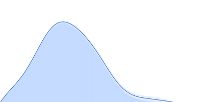
Inhibitor 6a was added to 1 mM concentration. The model displayed is a bead-occupancy and volume-corrected spatial representation derived from an aligned individual model cohort (damfilt) and does not correspond to the fit to the SAXS data displayed in this entry. Experimental temperature: UNKNOWN.
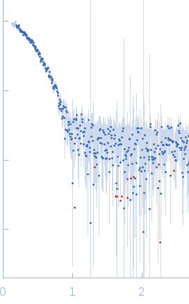
L-lactate dehydrogenase (PfMDH)
Mol. type   Protein
Organism   Plasmodium falciparum
Olig. state   Tetramer
Mon. MW   35.3 kDa
 
UniProt   Q6VVP7 (1-313)
Sequence   FASTA