A structural study of the cytoplasmic chaperone effect of 14-3-3 proteins on Ataxin-1.

Leysen S Jane Burnley R, Rodriguez E, Milroy LG, Soini L, Adamski CJ, Nitschke L, Davis R, Obsil T, Brunsveld L, Crabbe T, Yahya Zoghbi H, Ottmann C, Martin Davis J, J Mol Biol :167174 (2021) Europe PMC

SASDLT3 – Ataxin-1 AXH-C

Ataxin-1
MWexperimental 56 kDa
MWexpected 55 kDa
VPorod 90 nm3
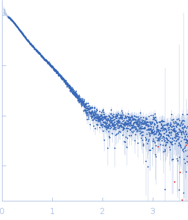
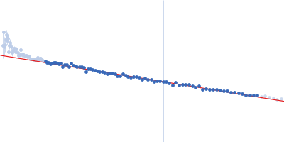
log I(s) 3.66×10⁻² 3.66×10⁻³ 3.66×10⁻⁴ 3.66×10⁻⁵
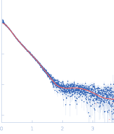
Ataxin-1 small angle scattering data  s, nm-1
ln I(s)
Ataxin-1 Guinier plot ln 3.66×10⁻² Rg: 4.3 nm 0 (4.3 nm)-2 s2
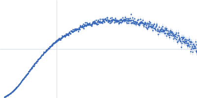
(sRg)2I(s)/I(0)
Ataxin-1 Kratky plot 1.104 0 3 sRg
p(r)
Ataxin-1 pair distance distribution function Rg: 4.4 nm 0 Dmax: 14.6 nm

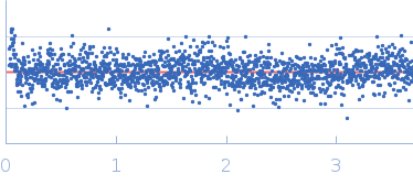
Data validation


Fits and models


log I(s)
 s, nm-1
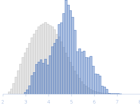
Ataxin-1 AXH-C Rg histogram Rg, nm
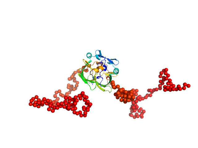
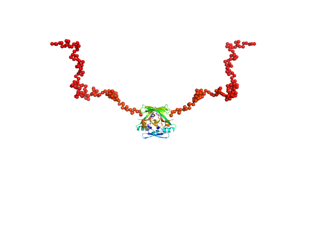
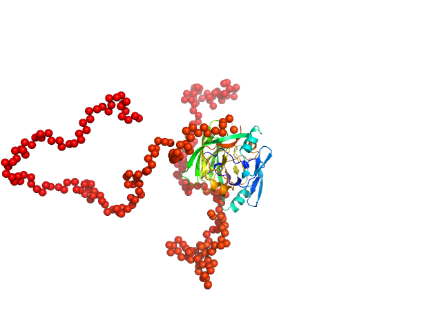
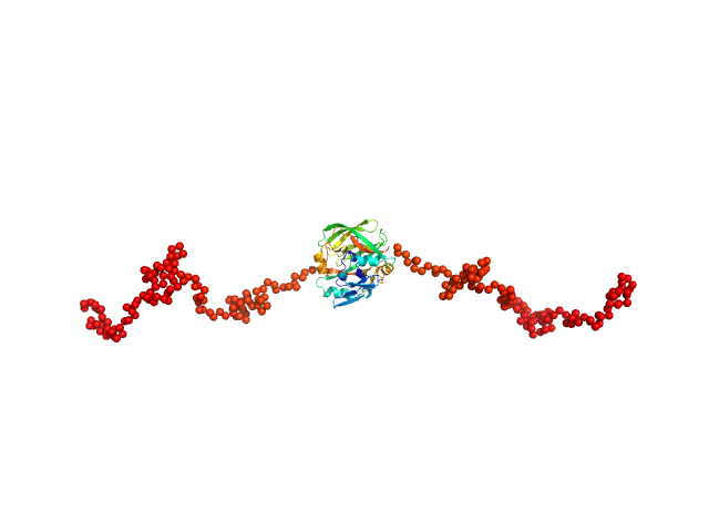
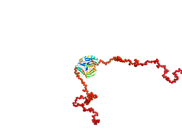
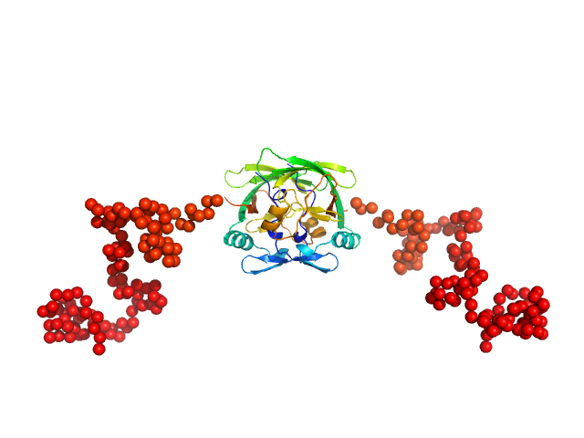
Ataxin-1 EOM/RANCH model

Synchrotron SAXS data from solutions of ataxin-1 AXH-C in 20 mM HEPES, 150 mM NaCl, 2 mM DTT, pH 7.5 were collected on the B21 beam line at the Diamond Light Source (Didcot, UK) using a Pilatus 2M detector at a wavelength of λ = 0.1 nm (I(s) vs s, where s = 4πsinθ/λ, and 2θ is the scattering angle). In-line size-exclusion chromatography (SEC) SAS was employed. The SEC parameters were as follows: A 45.00 μl sample at 12.8 mg/ml was injected onto a Shodex KW403 column at 25°C. 3 second data frames were collected through the SEC elution (620 data frames, total). The data were normalized to the intensity of the transmitted beam and radially averaged; the scattering of the solvent-blank was subtracted.

Storage temperature = UNKNOWN. Sample detector distance = UNKNOWN. Flow rate = UNKNOWN

Ataxin-1 (AXH-C)
Mol. type   Protein
Organism   Homo sapiens
Olig. state   Dimer
Mon. MW   27.7 kDa
 
UniProt   P54253 (562-815)
Sequence   FASTA