Kinetic and mechanistic characterization of Mycobacterium tuberculosis glutamyl-tRNA synthetase and determination of its oligomeric structure in solution

Paravisi S, Fumagalli G, Riva M, Morandi P, Morosi R, Konarev P, Petoukhov M Bernier S, Chênevert R, Svergun D, Curti B, Vanoni M, FEBS Journal 276(5):1398-1417 (2009) DOI

SASDLY5 – Monomer-dimer equilibrium of glutamyl-tRNA synthetase

Glutamate--tRNA ligase
MWexperimental 75 kDa
MWexpected 108 kDa
VPorod 123 nm3
log I(s) 2.36×10² 2.36×101 2.36×100 2.36×10⁻¹
Glutamate--tRNA ligase small angle scattering data  s, nm-1
ln I(s)
Glutamate--tRNA ligase Guinier plot ln 2.36×10² Rg: 3.5 nm 0 (3.5 nm)-2 s2
(sRg)2I(s)/I(0)
Glutamate--tRNA ligase Kratky plot 1.104 0 3 sRg
p(r)
Glutamate--tRNA ligase pair distance distribution function Rg: 3.6 nm 0 Dmax: 11 nm

Data validation


Fits and models


log I(s)
 s, nm-1
Glutamate--tRNA ligase PDB (PROTEIN DATA BANK) model
Glutamate--tRNA ligase PISA model

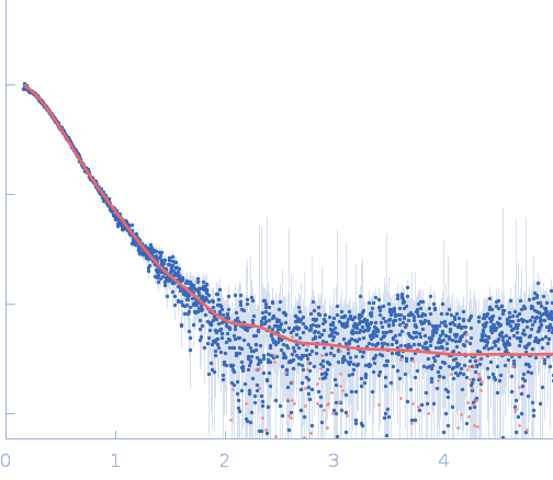
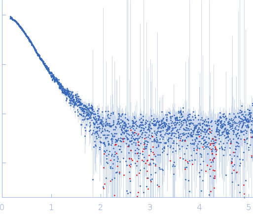
Synchrotron SAXS data from solutions of glutamyl-tRNA synthetase in 35 mM HEPES⁄NaOH, pH 6.5 buffer were collected on the EMBL X33 beam line at DORIS III (DESY, Hamburg, Germany) using a MAR 345 Image Plate detector at a sample-detector distance of 2.7 m and at a wavelength of λ = 0.15 nm (I(s) vs s, where s = 4πsinθ/λ, and 2θ is the scattering angle). One solute concentration of 2.00 mg/ml was measured. Two successive 60 second frames were collected. The data were normalized to the intensity of the transmitted beam and radially averaged; the scattering of the solvent-blank was subtracted.

Cell temperature = UNKNOWN. Storage temperature = UNKNOWN

Tags: X33
Glutamate--tRNA ligase (Gts)
Mol. type   Protein
Organism   Mycobacterium tuberculosis (strain ATCC 25618 / H37Rv)
Olig. state   Dimer
Mon. MW   53.9 kDa
 
UniProt   P9WFV9 (1-490)
Sequence   FASTA
 
PDB ID   1G59
 
PDB ID   1GTS