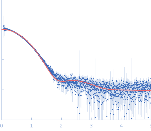
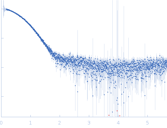
Nonstructural Proteins 7 and 8 of Feline Coronavirus Form a 2:1 Heterotrimer That Exhibits Primer-Independent RNA Polymerase Activity

Xiao Y, Ma Q, Restle T, Shang W, Svergun D, Ponnusamy R, Sczakiel G, Hilgenfeld R, Journal of Virology 86(8):4444-4454 (2012) DOI

SASDMJ9 – Plasmid pET11d- 229E710 encoding C-terminally His6-tagged HCoV-229E Nsp7-10 polyprotein

Replicase polyprotein 1a
MWexperimental 21 kDa
MWexpected 19 kDa
log I(s) 1.84×101 1.84×100 1.84×10⁻¹ 1.84×10⁻²
Replicase polyprotein 1a small angle scattering data  s, nm-1
ln I(s)
Replicase polyprotein 1a Guinier plot ln 1.84×101 Rg: 2.1 nm 0 (2.1 nm)-2 s2
(sRg)2I(s)/I(0)
Replicase polyprotein 1a Kratky plot 1.104 0 3 sRg

Data validation


Fits and models


log I(s)
 s, nm-1
Replicase polyprotein 1a PDB (PROTEIN DATA BANK) model

Synchrotron SAXS data from solutions of Plasmid pET11d- 229E710 encoding C-terminally His6-tagged HCoV-229E Nsp7-10 polyprotein in 10 mM Tris-HCl, 200 mM NaCl, 5 mM DTT, pH 7.5 were collected on the EMBL X33 beam line at the DORIS III, DESY storage ring (Hamburg, Germany) using a Pilatus 1M-W detector at a sample-detector distance of 2.7 m and at a wavelength of λ = 0.154 nm (I(s) vs s, where s = 4πsinθ/λ, and 2θ is the scattering angle). One solute concentration of 4.70 mg/ml was measured. Eight successive 15 second frames were collected. The data were normalized to the intensity of the transmitted beam and radially averaged; the scattering of the solvent-blank was subtracted.

Cell temperature = UNKNOWN. Storage temperature = UNKNOWN

Tags: X33
Replicase polyprotein 1a (Nsp7)
Mol. type   Protein
Organism   Severe acute respiratory syndrome-related coronavirus
Olig. state   Dimer
Mon. MW   9.3 kDa
 
UniProt   P0C6U8 (3837-3919)
Sequence   FASTA
 
PDB ID   7DCD