|
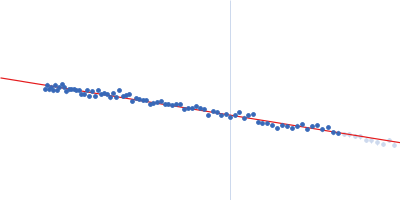
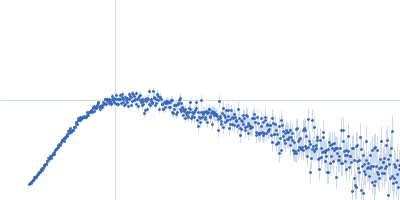
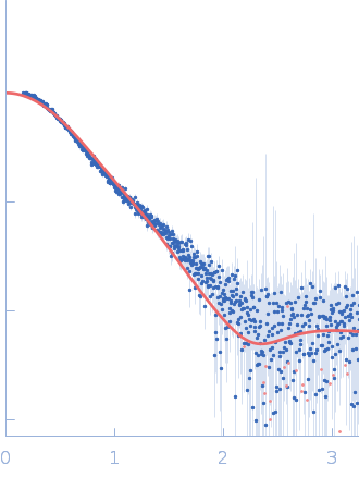
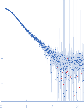
Synchrotron SAXS
data from solutions of
Listeria monocytogenes invasion protein internalin B
in
25mM Na-phosphate buffer, 150 mM NaCl, 1mM DTT, pH 7.5
were collected
on the
EMBL X33 beam line
at the DORIS III, DESY storage ring
(Hamburg, Germany)
using a 1D Gas detector detector
at a wavelength of λ = 0.15 nm
(I(s) vs s, where s = 4πsinθ/λ, and 2θ is the scattering angle).
One solute concentration of 32.00 mg/ml was measured.
15 successive
60 second frames were collected.
The data were normalized to the intensity of the transmitted beam and radially averaged; the scattering of the solvent-blank was subtracted.
Cell temperature = UNKNOWN. Storage temperature = UNKNOWN. Sample detector distance = UNKNOWN
|
|
Internalin B
(InlB)
|
| Mol. type |
|
Protein |
| Organism |
|
Listeria monocytogenes serotype 1/2a (strain EGD / Mackaness) |
| Olig. state |
|
Monomer |
| Mon. MW |
|
35.8 kDa |
| |
| UniProt |
|
P0DQD3
(1-320)
|
| Sequence |
|
FASTA |
| |
|
PDB ID
|
|
2UZX
|
| |
|
 s, nm-1