Comparative analysis of heparine oligosaccharides, heparin and heparan sulphate

Adriana Erica Miele.

SASDN54 – Heparin oligosaccharide DP12 in 50 mM Tris/HCl, 60 mM CaCl2, pH 7.4

Heparine oligosaccharide of 12 sugar units (6 disaccharides), digested by Heparinase I from purified HP glycosaminoglycan
MWI(0) 4 kDa
MWexpected 4 kDa
log I(s) 7.54×100 7.54×10⁻¹ 7.54×10⁻² 7.54×10⁻³
Heparine oligosaccharide of 12 sugar units (6 disaccharides), digested by Heparinase I from purified HP glycosaminoglycan small angle scattering data  s, nm-1
ln I(s)
Heparine oligosaccharide of 12 sugar units (6 disaccharides), digested by Heparinase I from purified HP glycosaminoglycan Guinier plot ln 7.55×100 Rg: 1.4 nm 0 (1.4 nm)-2 s2
(sRg)2I(s)/I(0)
Heparine oligosaccharide of 12 sugar units (6 disaccharides), digested by Heparinase I from purified HP glycosaminoglycan Kratky plot 1.104 0 3 sRg
p(r)
Heparine oligosaccharide of 12 sugar units (6 disaccharides), digested by Heparinase I from purified HP glycosaminoglycan pair distance distribution function Rg: 1.4 nm 0 Dmax: 8.6 nm

Data validation


There are no models related to this curve.

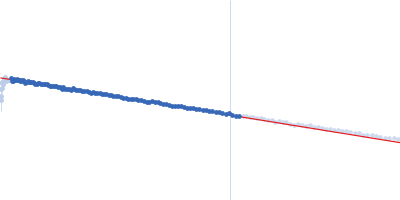
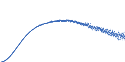
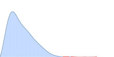
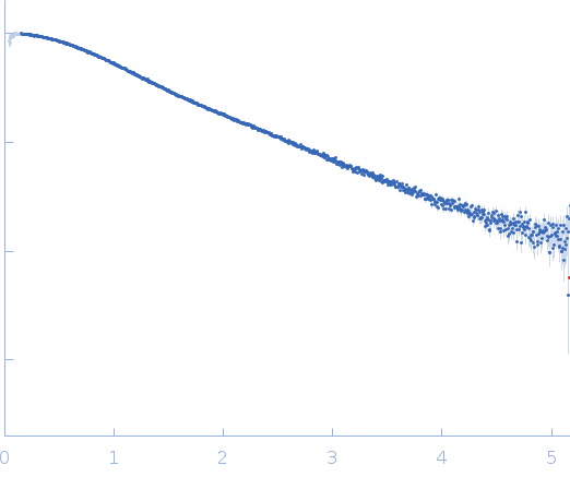
Synchrotron SAXS data from solutions of Heparin oligosaccharide DP12 in 50 mM Tris/HCl, 60 mM CaCl2, pH 7.4 were collected on the BM29 beam line at the ESRF storage ring (Grenoble, France) using a Pilatus3 2M detector at a sample-detector distance of 2 m and at a wavelength of λ = 0.1 nm (I(s) vs s, where s = 4πsinθ/λ, and 2θ is the scattering angle). One solute concentration of 5.00 mg/ml was measured at 20°C. 10 successive 0.500 second frames were collected. The data were normalized to the intensity of the transmitted beam and radially averaged; the scattering of the solvent-blank was subtracted.

Serial dilutions from 5 mg/ml to 1.25 mg/ml were collected under flow with the sample changer. At 60 mM Ca2+ aggregation started to appear. These oligosaccharides have been prepared by high resolution gel filtration of partial heparin lyase (Heparinase I) digestion of high quality porcin heparin. General formula* ∆HexA,2S - GlcNS,6S– (IdoUA,2S – GlcNS,6S)n n = number of disaccharide units. *Although the main disaccharide unit in these products is IdoUA,2S – GlcNS,6S, (approx 75%) saccharides in each size class show some variation in degree and pattern of sulphation. Uronic acid (HexA) at the non-reducing end of the oligosaccharides has a C4-C5 double bond as a result of the endolytic action of bacterial heparin lyase.

Heparine oligosaccharide of 12 sugar units (6 disaccharides), digested by Heparinase I from purified HP glycosaminoglycan (DP12)
Mol. type   Other
Organism   Sus scrofa domesticus
Olig. state   Monomer
Mon. MW   3.9 kDa
Chemical formula