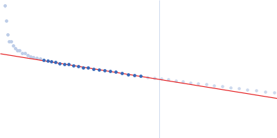
Synchrotron SAXS data from solutions of Heparin oligosaccharide in 50 mM Tris/HCl, pH 7.4 were collected on the SWING beam line at the SOLEIL storage ring (Saint-Aubin, France) using a Eiger 4M detector at a sample-detector distance of 2 m and at a wavelength of λ = 10.33 nm (I(s) vs s, where s = 4πsinθ/λ, and 2θ is the scattering angle). One solute concentration of 2.50 mg/ml was measured at 20°C. 20 successive 0.500 second frames were collected. The data were normalized to the intensity of the transmitted beam and radially averaged; the scattering of the solvent-blank was subtracted.
High grade unfractionated Heparin is a glycosaminoglycan polymer with a high degree of sulfation. Although the main disaccharide in heparin is the trisulphated unit GlcNS,6S – IdoA,2S heparin also contains di-, mono- and non-sulphated units; thus each size class of oligosaccharide, though substantially homogeneous in molecular size, contains structures that vary in content and pattern of sulphation.
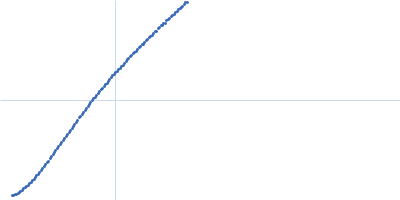
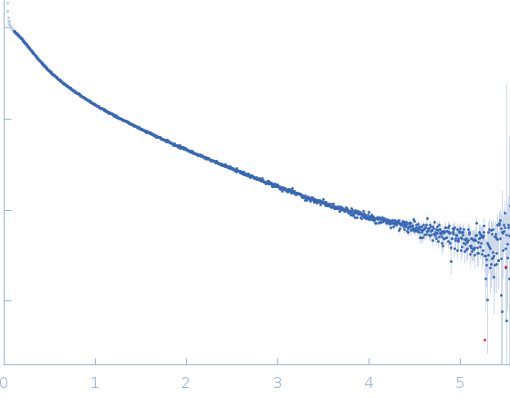 s, nm-1