LARGE1 processively polymerizes length-controlled matriglycan on prodystroglycan.

Joseph S, Schnicker NJ, Spellmon N, Xu Z Yan R, Yu Z, Davulcu O, Yang T, Hopkins J, Anderson ME, Venzke D, Campbell KP, Nat Commun 16(1):9028 (2025) Europe PMC

SASDNG8 – LARGE xylosyl- and glucuronyltransferase 1 (LARGE1dTM) dimer treated with PNGase F enzyme

Xylosyl- and glucuronyltransferase LARGE1
MWexperimental 207 kDa
MWexpected 179 kDa
log I(s) 1.58×100 1.58×10⁻¹ 1.58×10⁻² 1.58×10⁻³
Xylosyl- and glucuronyltransferase LARGE1 small angle scattering data  s, nm-1
ln I(s)
Xylosyl- and glucuronyltransferase LARGE1 Guinier plot ln 1.58×100 Rg: 4.3 nm 0 (4.3 nm)-2 s2
(sRg)2I(s)/I(0)
Xylosyl- and glucuronyltransferase LARGE1 Kratky plot 1.104 0 3 sRg
p(r)
Xylosyl- and glucuronyltransferase LARGE1 pair distance distribution function Rg: 4.4 nm 0 Dmax: 18 nm

Data validation


Fits and models


log I(s)
 s, nm-1
Xylosyl- and glucuronyltransferase LARGE1 DAMMIF model

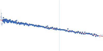
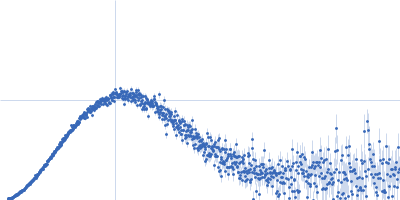
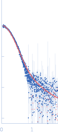
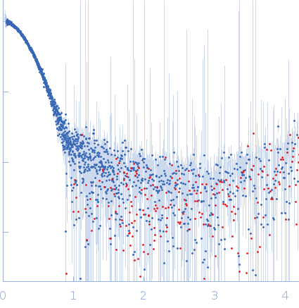
Synchrotron SAXS data from solutions of PNGase F treated LARGE1dTM in buffer (20 mM HEPES pH 7.4, 150 mM NaCl) were collected on the BioCAT 18-ID-D beamline at the Advanced Photon Source (APS) (Chicago, IL, USA) using a Eiger2 XE 9M detector at a sample-detector distance of 3.67 m and at a wavelength of λ = 0.1033 nm (I(s) vs s, where s = 4πsinθ/λ, and 2θ is the scattering angle). In-line size-exclusion chromatography (SEC)-MALS-SAXS was employed. The SEC parameters were as follows: A 500 μl sample at 1 mg/ml was injected at a 0.6 ml/min flow rate onto a Superdex 200 increase 10/300 GL column (GE healthcare) at 23°C. 2500 successive 1 second frames were collected. The data were normalized to the intensity of the transmitted beam and radially averaged; the scattering of the solvent-blank was subtracted.

Xylosyl- and glucuronyltransferase LARGE1 (LARGE1)
Mol. type   Protein
Organism   Homo sapiens
Olig. state   Dimer
Mon. MW   89.7 kDa
 
UniProt   O95461 (34-756)
Sequence   FASTA