Solution structure of the type I polyketide synthase Pks13 from Mycobacterium tuberculosis.

Bon C, Cabantous S, Julien S, Guillet V, Chalut C, Rima J, Brison Y, Malaga W, Sanchez-Dafun A, Gavalda S, Quémard A, Marcoux J, Waldo GS, Guilhot C, Mourey L, BMC Biol 20(1):147 (2022) Europe PMC

SASDNR9 – Subfragment ACP1KSAT tridomain of polyketide synthase Pks13 at neutral pH

Polyketide synthase Pks13
MWI(0) 116 kDa
MWexpected 116 kDa
VPorod 186 nm3
log I(s) 3.05×10² 3.05×101 3.05×100 3.05×10⁻¹
Polyketide synthase Pks13 small angle scattering data  s, nm-1
ln I(s)
Polyketide synthase Pks13 Guinier plot ln 3.05×10² Rg: 3.8 nm 0 (3.8 nm)-2 s2
(sRg)2I(s)/I(0)
Polyketide synthase Pks13 Kratky plot 1.104 0 3 sRg
p(r)
Polyketide synthase Pks13 pair distance distribution function Rg: 4.0 nm 0 Dmax: 12.9 nm

Data validation


Fits and models


log I(s)
 s, nm-1
Polyketide synthase Pks13 DAMMIN model

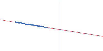
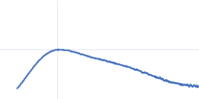
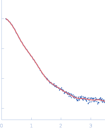
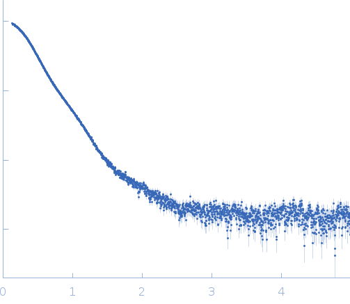
Synchrotron SAXS data from solutions of the sub-fragment ACP1KSAT tridomain of polyketide synthase Pks13 at neutral pH in 50 mM Tris-HCl 50 mM NaCl, pH 8.5 were collected on the EMBL X33 beam line at the DORIS III storage ring (Hamburg, Germany) using a MAR 345 Image Plate detector at a wavelength of λ = 0.15 nm (I(s) vs s, where s = 4πsinθ/λ, and 2θ is the scattering angle). One solute concentration of 5.70 mg/ml was measured at 12°C. The data were normalized to the intensity of the transmitted beam and radially averaged; the scattering of the solvent-blank was subtracted.

Sample detector distance = UNKNOWN. Number of frames = UNKNOWN

Tags: X33
Polyketide synthase Pks13 (fACP1-KS-AT)
Mol. type   Protein
Organism   Mycobacterium tuberculosis (strain ATCC 25618 / H37Rv)
Olig. state   Monomer
Mon. MW   115.8 kDa
 
UniProt   I6X8D2 (1-1063)
Sequence   FASTA