Structural insights into 3Fe–4S ferredoxins diversity in M. tuberculosis highlighted by a first redox complex with P450

Gilep A, Varaksa T, Bukhdruker S, Kavaleuski A, Ryzhykau Y, Smolskaya S, Sushko T, Tsumoto K, Grabovec I, Kapranov I, Okhrimenko I, Marin E, Shevtsov M, Mishin A, Kovalev K, Kuklin A, Gordeliy V, Kaluzhskiy L, Gnedenko O, Yablokov E, Ivanov A, Borshchevskiy V, Strushkevich N, Frontiers in Molecular Biosciences 9 (2023) DOI

SASDPL2 – Fused complex of cytochrome P450 143 and ferredoxin Rv1786 (Cyp143 and rv1786)

His-tagged fused complex of cytochrome P450 143 and ferredoxin Rv1786
MWexperimental 52 kDa
MWexpected 54 kDa
VPorod 85 nm3
log I(s) 8.12×10⁻² 8.12×10⁻³ 8.12×10⁻⁴ 8.12×10⁻⁵
His-tagged fused complex of cytochrome P450 143 and ferredoxin Rv1786 small angle scattering data  s, nm-1
ln I(s)
His-tagged fused complex of cytochrome P450 143 and ferredoxin Rv1786 Guinier plot ln 8.12×10⁻² Rg: 2.5 nm 0 (2.5 nm)-2 s2
(sRg)2I(s)/I(0)
His-tagged fused complex of cytochrome P450 143 and ferredoxin Rv1786 Kratky plot 1.104 0 3 sRg
p(r)
His-tagged fused complex of cytochrome P450 143 and ferredoxin Rv1786 pair distance distribution function Rg: 2.5 nm 0 Dmax: 9 nm

Data validation


Fits and models


log I(s)
 s, nm-1
Fused complex of cytochrome P450 143 and ferredoxin Rv1786 (Cyp143 and rv1786) Rg histogram Rg, nm
His-tagged fused complex of cytochrome P450 143 and ferredoxin Rv1786 EOM/RANCH model

log I(s)
 s, nm-1
His-tagged fused complex of cytochrome P450 143 and ferredoxin Rv1786 CORAL model

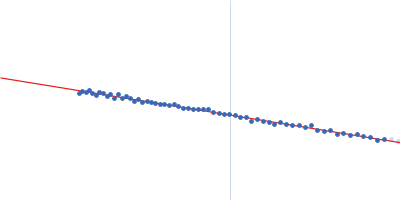
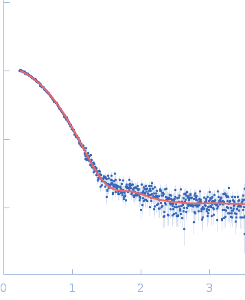
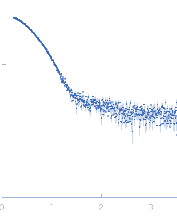
Synchrotron SAXS data from solutions of a fused complex between cytochrome P450 143 and ferredoxin Rv1786 in 300 mM NaCl, 50 mM Tris/TrisHCl, 10% glycerol, pH 7.4 were collected on the BM29 beam line at the ESRF (Grenoble, France) using a Pilatus 1M detector at a sample-detector distance of 2.9 m and at a wavelength of λ = 0.09918 nm (I(s) vs s, where s = 4πsinθ/λ, and 2θ is the scattering angle). One solute concentration of 2.20 mg/ml was measured at 20°C. 16 successive 0.500 second frames were collected. The data were normalized to the intensity of the transmitted beam and radially averaged; the scattering of the solvent-blank was subtracted.

His-tagged fused complex of putative cytochrome P450 143 (UniProt ID: P9WPL3, https://www.uniprot.org/uniprot/P9WPL3) and probable ferredoxin Rv1786 (UniProt ID: O53937, https://www.uniprot.org/uniprot/O53937) from Mycobacterium tuberculosis expressed in E. coli.

His-tagged fused complex of cytochrome P450 143 and ferredoxin Rv1786 (CYP143-Rv1786)
Mol. type   Protein
Organism   Mycobacterium tuberculosis
Olig. state   Monomer
Mon. MW   53.6 kDa
Sequence   FASTA