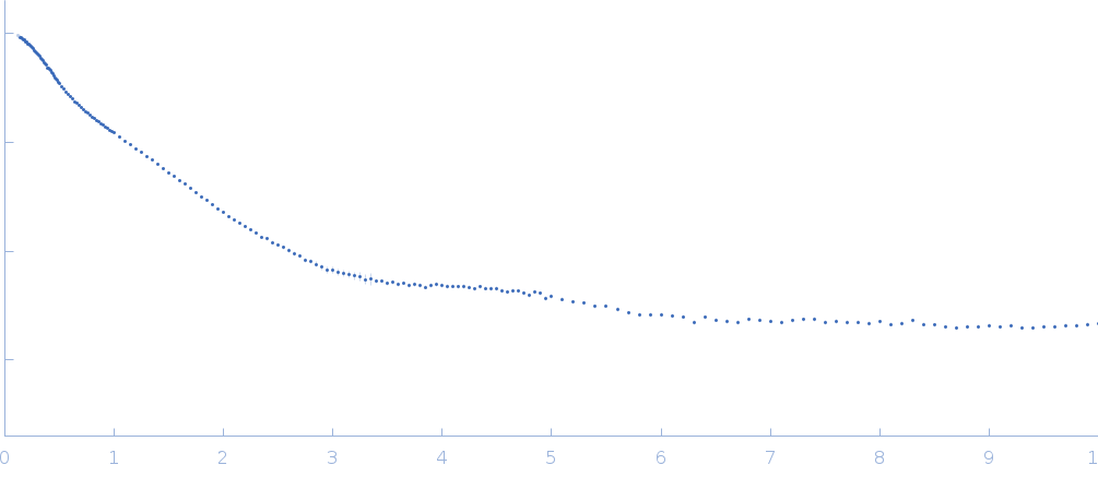
Synchrotron SAXS data from solutions of MHV coronavirus putative packaging signal RNA in 50 mM NaCl, 50 μM EDTA, 10 mM MOPS, pH 7 were collected on the 16-ID (LiX) beam line at the National Synchrotron Light Source II (NSLS-II; Upton, NY, USA) using a Pilatus3 S 1M detector at a wavelength of λ = 0.0918 nm (I(s) vs s, where s = 4πsinθ/λ, and 2θ is the scattering angle). Solute concentrations ranging between 0.9 and 3 mg/ml were measured at 10°C. Five successive 1 second frames were collected. The data were normalized to the intensity of the transmitted beam and radially averaged; the scattering of the solvent-blank was subtracted.
Data collected from two detector positions were merged to obtain the SAXS/WAXS profile.
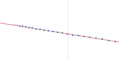
 s, nm-1
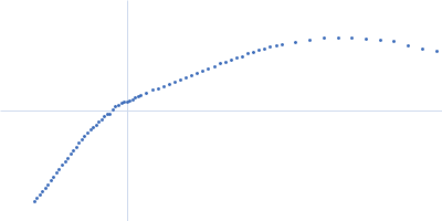