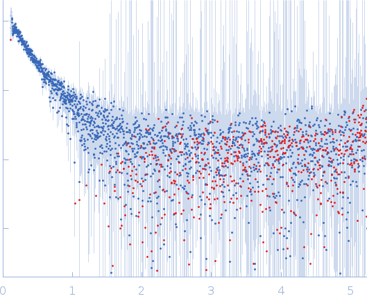
Synchrotron SAXS data from solutions of MHV coronavirus nucleocapsid protein N in 70 mM KCl, 50 μM EDTA, 25 mM HEPES, pH 7 were collected on the G1 beam line at the Cornell High Energy Synchrotron Source (CHESS; Ithaca, NY, USA) using a Eiger 4M detector at a sample-detector distance of 1.6 m and at a wavelength of λ = 0.1265 nm (I(s) vs s, where s = 4πsinθ/λ, and 2θ is the scattering angle). Solute concentrations ranging between 0.9 and 0.5 mg/ml were measured at 22°C. 20 successive 1 second frames were collected. The data were normalized to the intensity of the transmitted beam and radially averaged; the scattering of the solvent-blank was subtracted.
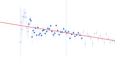
 s, nm-1
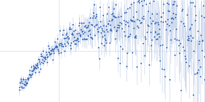