Molecular Determinants of Fibrillation in a Viral Amyloidogenic Domain from Combined Biochemical and Biophysical Studies

Nilsson J Baroudi H, Gondelaud F, Pesce G, Bignon C, Ptchelkine D, Chamieh J, Cottet H, Kajava A, Longhi S, International Journal of Molecular Sciences 24(1):399 (2022) DOI

SASDQC7 – Region within the N-terminal domain of P/V/W protein of Hendra virus (PNT3 region) mutated YYY --> AAA

Non-structural protein V (Y211A, Y212A, Y213A mutant)
MWexperimental 21 kDa
MWexpected 15 kDa
VPorod 49 nm3
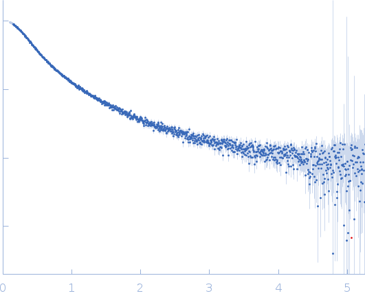
log I(s) 2.14×10⁻² 2.14×10⁻³ 2.14×10⁻⁴ 2.14×10⁻⁵
Non-structural protein V (Y211A, Y212A, Y213A mutant) small angle scattering data  s, nm-1
ln I(s)
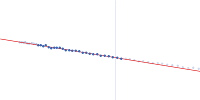
Non-structural protein V (Y211A, Y212A, Y213A mutant) Guinier plot ln 2.15×10⁻² Rg: 4.0 nm 0 (4.0 nm)-2 s2
(sRg)2I(s)/I(0)
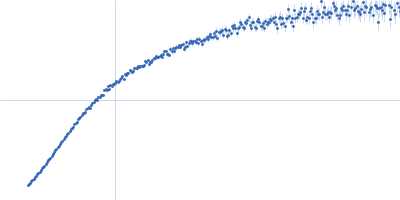
Non-structural protein V (Y211A, Y212A, Y213A mutant) Kratky plot 1.104 0 3 sRg
Dmax: 19 nm

Data validation


There are no models related to this curve.

Synchrotron SAXS data from solutions of the PNT3 region of P/V/W protein (mutated YYY --> AAA) in 50 mM sodium phosphate, pH 7.2 were collected on the SWING beam line at SOLEIL (Saint-Aubin, France) using a Eiger 4M detector at a sample-detector distance of 2 m and at a wavelength of λ = 0.10332 nm (I(s) vs s, where s = 4πsinθ/λ, and 2θ is the scattering angle). In-line size-exclusion chromatography (SEC) SAS was employed. The SEC parameters were as follows: A 50.00 μl sample at 5 mg/ml was injected at a 0.30 ml/min flow rate onto a Agilent AdvanceBio SEC 2.7 µm - 130 Å column at 20°C. 810 successive 0.990 second frames were collected. The data were normalized to the intensity of the transmitted beam and radially averaged; the scattering of the solvent-blank was subtracted.

Tags: idp
Non-structural protein V (Y211A, Y212A, Y213A mutant) (HeV_PNT3(AAA))
Mol. type   Protein
Organism   Hendra virus (isolate Horse/Autralia/Hendra/1994)
Olig. state   Monomer
Mon. MW   14.9 kDa
 
UniProt   O55777 (200-310)
Sequence   FASTA