Visualizing RNA conformational and architectural heterogeneity in solution.

Ding J, Lee YT, Bhandari Y, Schwieters CD, Fan L, Yu P, Tarosov SG, Stagno JR, Ma B, Nussinov R, Rein A, Zhang J, Wang YX, Nat Commun 14(1):714 (2023) Europe PMC

SASDQH7 – Thermoanearobacter tengcongensis (Tte) fecB RNA riboswitch aptamer domain (rCbl) in the presence of coenzyme B12

Thermoanearobacter tengcongensis (Tte) fecB riboswitch aptamer domain
MWexperimental 98 kDa
MWexpected 68 kDa
VPorod 230 nm3
log I(s) 3.86×10⁻² 3.86×10⁻³ 3.86×10⁻⁴ 3.86×10⁻⁵
Thermoanearobacter tengcongensis (Tte) fecB riboswitch aptamer domain small angle scattering data  s, nm-1
ln I(s)
Thermoanearobacter tengcongensis (Tte) fecB riboswitch aptamer domain Guinier plot ln 3.87×10⁻² Rg: 5.7 nm 0 (5.7 nm)-2 s2
(sRg)2I(s)/I(0)
Thermoanearobacter tengcongensis (Tte) fecB riboswitch aptamer domain Kratky plot 1.104 0 3 sRg
Dmax: 19.7 nm

Data validation


Fits and models


log I(s)
 s, nm-1
Thermoanearobacter tengcongensis (Tte) fecB riboswitch aptamer domain OTHER model

log I(s)
 s, nm-1
Thermoanearobacter tengcongensis (Tte) fecB riboswitch aptamer domain OTHER model

log I(s)
 s, nm-1
Thermoanearobacter tengcongensis (Tte) fecB riboswitch aptamer domain OTHER model

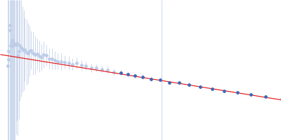
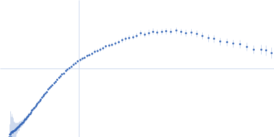
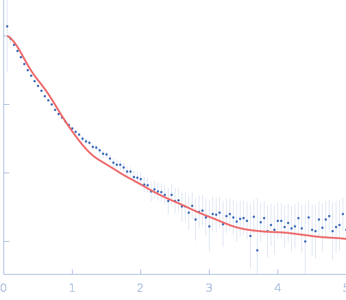
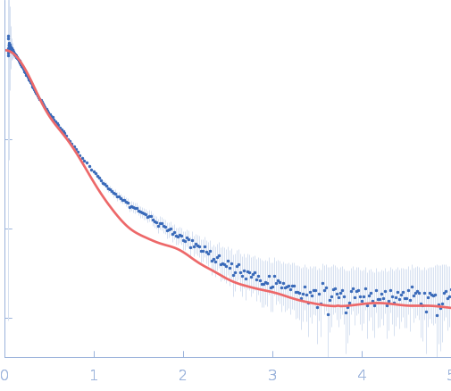
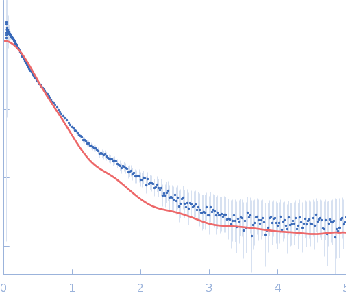
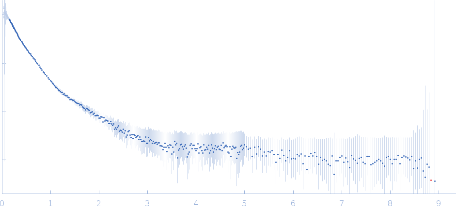
Synchrotron SAXS data from solutions of the fecB RNA riboswitch aptamer domain (rCbl) in 50 mM MES pH 6.0, 10 mM KCl, 1 mM MgCl2, 4-18 μM coenzyme B12 ligand, were collected on the 12ID-B SAXS/WAXS beam line at the Advanced Photon Source (APS; Argonne National Laboratory; Lemont, IL, USA) using a Pilatus 2M detector at a sample-detector distance of 1.9 m and at a wavelength of λ = 0.0932 nm (I(s) vs s, where s = 4πsinθ/λ, and 2θ is the scattering angle). One solute concentration of 0.07 mg/ml was measured at 10°C. 225 successive 1 second frames were collected. The data were normalized to the intensity of the transmitted beam and radially averaged; the scattering of the solvent-blank was subtracted.

AFM single-particle visualization of rCbl in presence of ligands revealed a mixture of monomeric and dimeric species, each with various conformations. A few higher order oligomers were also observed. See the manuscript for detailed description of conforrmational and structural heterogeneity. The non- linearity of Guinier plot confirms this is not a monodisperse system. The Rg value may depend on the selection of the Guinier region.

Thermoanearobacter tengcongensis (Tte) fecB riboswitch aptamer domain (rCbl with ligand)
Mol. type   RNA
Organism   marine metagenome
Olig. state   Other
Mon. MW   68.3 kDa
Sequence   FASTA