Visualization of conformational transition of GRP94 in solution.

Sun S, Zhu R, Zhu M, Wang Q, Li N, Yang B, Life Sci Alliance 7(2) (2024) Europe PMC

SASDQH9 – GRP94 protein (Endoplasmin) bound to AMP-PNP

Endoplasmin
MWexperimental 153 kDa
MWexpected 169 kDa
VPorod 260 nm3
log I(s) 5.86×101 5.86×100 5.86×10⁻¹ 5.86×10⁻²
Endoplasmin small angle scattering data  s, nm-1
ln I(s)
Endoplasmin Guinier plot ln 5.86×101 Rg: 5.5 nm 0 (5.5 nm)-2 s2
(sRg)2I(s)/I(0)
Endoplasmin Kratky plot 1.104 0 3 sRg
Dmax: 24.5 nm

Data validation


There are no models related to this curve.

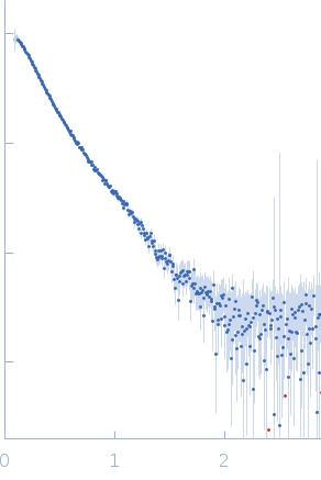
Synchrotron SAXS data from solutions of GRP94 protein bound to AMP-PNP in 25 mM HEPES, 200 mM NaCl, 1 mM TCEP, pH 8 were collected on the BL19U2 beam line at the Shanghai Synchrotron Radiation Facility, (SSRF, Shanghai, China) using a Pilatus 1 M detector detector at a sample-detector distance of 2.8 m and at a wavelength of λ = 0.103 nm (I(s) vs s, where s = 4πsinθ/λ, and 2θ is the scattering angle). In-line size-exclusion chromatography (SEC) SAS was employed. The SEC parameters were as follows: A 100.00 μl sample at 10 mg/ml was injected at a 0.50 ml/min flow rate onto a Wyatt WTC-030S5 5μm 300+, 7.8x300 mm column at 25°C. 720 successive 1 second frames were collected. The data were normalized to the intensity of the transmitted beam and radially averaged; the scattering of the solvent-blank was subtracted.

AMP-PNP = Phosphoaminophosphonic acid-adenylate ester, https://pubchem.ncbi.nlm.nih.gov/compound/Phosphoaminophosphonic-acid-adenylate-ester

Endoplasmin (GRP94)
Mol. type   Protein
Organism   Mus musculus
Olig. state   Dimer
Mon. MW   84.6 kDa
 
UniProt   P08113 (22-754)
Sequence   FASTA