|
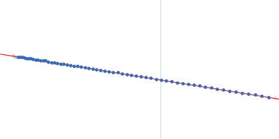
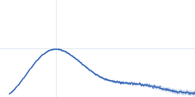
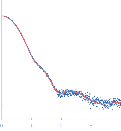
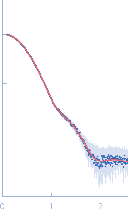
Synchrotron SAXS data from solutions of Endo-D-arabinanase,20 mM Tris-HCl, 200 mM NaCl, pH 7.5 were collected on the BL-10C beam line at the Photon Factory (PF), High Energy Accelerator Research Organization (KEK; Tsukuba, Japan) using a Pilatus3 2M detector at a sample-detector distance of 2.1 m and at a wavelength of λ = 0.1 nm (I(s) vs s, where s = 4πsinθ/λ, and 2θ is the scattering angle). In-line size-exclusion chromatography (SEC) SAS was employed. The SEC parameters were as follows: A 130.00 μl sample at 2.4 mg/ml was injected at a 0.02 ml/min flow rate onto a Shodex KW403 column at 20°C. 247 successive 20 second frames were collected. The data were normalized to the intensity of the transmitted beam and radially averaged; the scattering of the solvent-blank was subtracted.
|
|
Endo-D-arabinanase
(EndoMA1)
|
| Mol. type |
|
Protein |
| Organism |
|
Microbacterium arabinogalactanolyticum |
| Olig. state |
|
Dimer |
| Mon. MW |
|
53.7 kDa |
| Sequence |
|
FASTA |
| |
|
PDB ID
|
|
8HHV
|
| |
|
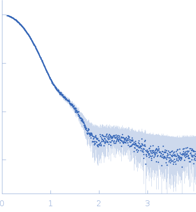 s, nm-1