Identification of structural determinants of nicotinamide phosphoribosyl transferase (NAMPT) activity and substrate selectivity.

Houry D, Raasakka A, Ferrario E, Niere M, Bifulco E, Kursula P, Ziegler M, J Struct Biol :108004 (2023) Europe PMC

SASDR58 – Human nicotinamide phosphoribosyltransferase Δ42-51 loop mutant (NAMPT Δ42-51)

Nicotinamide phosphoribosyltransferase Δ42-51
MWI(0) 73 kDa
MWexpected 111 kDa
VPorod 159 nm3
log I(s) 1.14×10⁴ 1.14×10³ 1.14×10² 1.14×101
Nicotinamide phosphoribosyltransferase Δ42-51 small angle scattering data  s, nm-1
ln I(s)
Nicotinamide phosphoribosyltransferase Δ42-51 Guinier plot ln 1.14×10⁴ Rg: 3.2 nm 0 (3.2 nm)-2 s2
(sRg)2I(s)/I(0)
Nicotinamide phosphoribosyltransferase Δ42-51 Kratky plot 1.104 0 3 sRg
p(r)
Nicotinamide phosphoribosyltransferase Δ42-51 pair distance distribution function Rg: 3.3 nm 0 Dmax: 11.0 nm

Data validation


There are no models related to this curve.

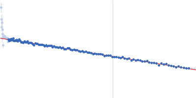
Synchrotron SAXS data from solutions of NAMPT Δ42-51 in 20 mM Tris-HCl, 500 mM NaCl, 6 mM MgCl2, 5% (v/v) glycerol, pH 8 were collected on the EMBL P12 beam line at PETRA III (DESY, Hamburg, Germany) using a Pilatus 6M detector at a sample-detector distance of 3 m and at a wavelength of λ = 0.124 nm (I(s) vs s, where s = 4πsinθ/λ, and 2θ is the scattering angle). One solute concentration of 1.86 mg/ml was measured at 10°C. 40 successive 0.045 second frames were collected. The data were normalized to the intensity of the transmitted beam and radially averaged; the scattering of the solvent-blank was subtracted.

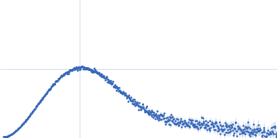
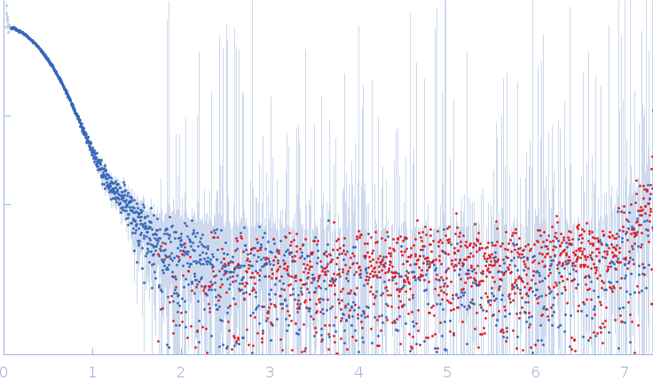
Dimeric His-tagged human NAMPT Δ42-51 mutant without ligands.

Nicotinamide phosphoribosyltransferase Δ42-51 (NAMPT Δ42-51)
Mol. type   Protein
Organism   Homo sapiens
Olig. state   Dimer
Mon. MW   55.7 kDa
 
UniProt   P43490 (2-491)
Sequence   FASTA