Synchrotron SAXS
data from solutions of
M1-linked tetraubiquitin, M1(1-73)-Ub4
in
20 mM sodium phosphate, 0.5 mM EDTA, 0.02 % NaN3, pH 6.8
were collected
on the
BioCAT 18ID beam line
at the Advanced Photon Source (APS), Argonne National Laboratory storage ring
(Lemont, IL, USA)
using a Pilatus3 X 1M detector
at a sample-detector distance of 3.7 m and
at a wavelength of λ = 0.1033 nm
(I(s) vs s, where s = 4πsinθ/λ, and 2θ is the scattering angle).
One solute concentration of 7.19 mg/ml was measured
at 20°C.
21 successive
0.500 second frames were collected.
The data were normalized to the intensity of the transmitted beam and radially averaged; the scattering of the solvent-blank was subtracted.
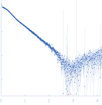 s, nm-1