|
SEC-SAXS data were collected at beamline B21, Diamond Light Source (Didcot, UK) that operates at a wavelength of 0.1 nm at a photon flux of ~10¹² photons per second and features a fixed camera length of 4.014 m. It is equipped with an Agilent 1200 HPLC system. Tag-free chicken Netrin-1 ΔC was shipped to the beamline at 1.0 mg/ml in 50 mM tris, pH 7.5, 1.0 M NaCl. Using a PD-10 desalting column with Sephadex G-25 resin, the buffer was changed to 50 mM tris, pH 7.5, 0.2 M NaCl and the protein was then concentrated to 9.23 ± 0.09 mg/ml (186 ± 2 µM) using an Amicon-4 concentrator with 30 kDa molecular weight cut-off. 20 µl of heparin oligosaccharide dp8 (10 mg/ml in water) was added to 100 µl protein solution and let to incubate for 40 - 80 min before injection.The sample was then centrifuged at 13000 rpm in an Eppendorf MiniSpin® centrifuge followed by filtration through a 0.1 µM Amicon Ultrafree-MC spin filter. 50 µl of sample were injected without further delay into the buffer equilibrated 4.6 ml Shodex KW 403-4F size exclusion column from where the protein eluted through the diffraction flow cell at a rate of 0.16 ml/min. X-ray images were collected with 3 s exposure time.
Experiment ID: sm16028-7/379532. Additional modelling information, including log files, are provided in the full entry zip archive. Each respective fit displays two maps: 1) The refined averaged map (top) and; 2) The corresponding object support (bottom).
|
|
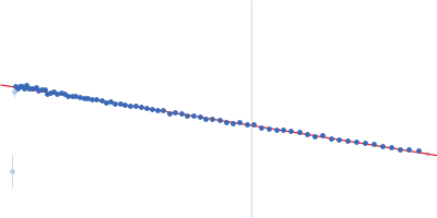
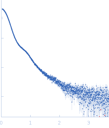 s, nm-1
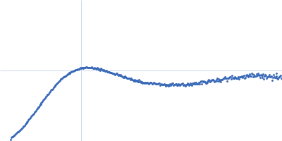