| MWexperimental | 585 | kDa |
| MWexpected | 264 | kDa |
| VPorod | 767 | nm3 |
|
log I(s)
5.23×100
5.23×10⁻¹
5.23×10⁻²
5.23×10⁻³
|
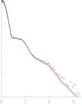
![Sulfite reductase [NADPH] flavoprotein alpha-component small angle scattering data](/media/intensities_files/scattering_plots/SASDS22_dat_img.png) s, nm-1
s, nm-1
|
|
|
|
![Sulfite reductase [NADPH] flavoprotein alpha-component SASREF model](/media/pdb_file/images/SASDS22_fit2_model1_img.png "Load 3D view")
|
|
|
|
SANS data from solutions of sulfite reductase flavoprotein crosslinked octamer in 50 mM KPi, 100 mM NaCl, 1 mM EDTA, pH 7.8 were collected using the EQ-SANS (BL-6) spallation neutron source (Oak Ridge, Tennessee, USA). This sample is an octamer of His-tagged sulfite reductase flavoprotein crosslinked subunits. Three instrument configurations were used: 9 m sample-to-detector distance with 1.5 nm wavelength band, 4 m sample-to-detector distance with 0.6 nm wavelength band, and 1.3 m sample-to-detector distance with 0.4 nm wavelength band. Sample concentration: 7 mg/mL. Neutron exposure time: 3 hours / instrument configuration. Sample temperature: 8°C.
# |
|
|||||||||||||||||||||||||||
![Sulfite reductase [NADPH] flavoprotein alpha-component Guinier plot](/media//intensities_files/scattering_plots/SASDS22_guinier_img.png)
![Sulfite reductase [NADPH] flavoprotein alpha-component Kratky plot](/media/intensities_files/scattering_plots/SASDS22_kratky_img.png)
![Sulfite reductase [NADPH] flavoprotein alpha-component pair distance distribution function](/media/p_of_R_files/pofr_images/SASDS22_pofr_img.png)