Structural properties of the HNF-1A transactivation domain

Kind L, Driver M, Raasakka A, Onck P, Njølstad P, Arnesen T, Kursula P, Frontiers in Molecular Biosciences 10 (2023) DOI

SASDS29 – The DNA binding and transactivation domains (DBD-TAD) of Hepatocyte nuclear factor 1-alpha (HNF-1A)

Hepatocyte nuclear factor 1-alpha
MWexperimental 54 kDa
MWexpected 62 kDa
VPorod 109 nm3
log I(s) 3.90×10⁻² 3.90×10⁻³ 3.90×10⁻⁴ 3.90×10⁻⁵
Hepatocyte nuclear factor 1-alpha small angle scattering data  s, nm-1
ln I(s)
Hepatocyte nuclear factor 1-alpha Guinier plot ln 3.91×10⁻² Rg: 4.9 nm 0 (4.9 nm)-2 s2
(sRg)2I(s)/I(0)
Hepatocyte nuclear factor 1-alpha Kratky plot 1.104 0 3 sRg
p(r)
Hepatocyte nuclear factor 1-alpha pair distance distribution function Rg: 4.8 nm 0 Dmax: 17 nm

Data validation


There are no models related to this curve.

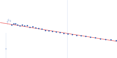
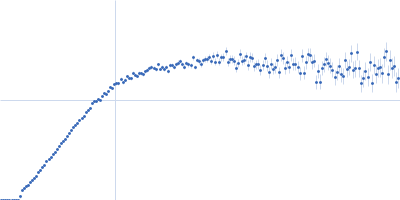
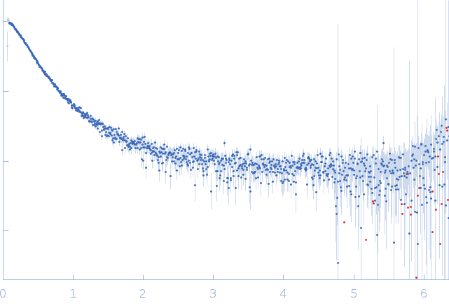
Synchrotron SAXS data from solutions of the DNA binding and transactivation domains of hepatocyte nuclear factor 1-alpha in 4 mM Tris, 100 mM NaCl, 1 mM TCEP, pH 8.5 were collected on the CoSAXS beam line at MAX IV (Lund, Sweden) using a Eiger2 4M detector at a wavelength of λ = 0.1 nm (I(s) vs s, where s = 4πsinθ/λ, and 2θ is the scattering angle). One solute concentration of 2.50 mg/ml was measured at 10°C. 300 successive 0.020 second frames were collected. The data were normalized to the intensity of the transmitted beam and radially averaged; the scattering of the solvent-blank was subtracted.

Sample detector distance = UNKNOWN

Hepatocyte nuclear factor 1-alpha (HNF-1A DBD-TAD)
Mol. type   Protein
Organism   Homo sapiens
Olig. state   Monomer
Mon. MW   62.2 kDa
 
UniProt   P20823 (83-631)
Sequence   FASTA