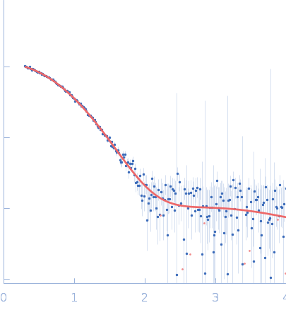
Illuminating the mechanism and allosteric behavior of NanoLuc luciferase.

Nemergut M, Pluskal D, Horackova J, Sustrova T, Tulis J, Barta T, Baatallah R, Gagnot G, Novakova V, Majerova M, Sedlackova K, Marques SM, Toul M, Damborsky J, Prokop Z, Bednar D, Janin YL, Marek M, Nat Commun 14(1):7864 (2023) Europe PMC

SASDSQ9 – NanoLuc luciferase

Oplophorus-luciferin 2-monooxygenase catalytic subunit
MWexperimental 20 kDa
MWexpected 20 kDa
VPorod 38 nm3
log I(s) 4.97×10⁻¹ 4.97×10⁻² 4.97×10⁻³ 4.97×10⁻⁴
Oplophorus-luciferin 2-monooxygenase catalytic subunit small angle scattering data  s, nm-1
ln I(s)
Oplophorus-luciferin 2-monooxygenase catalytic subunit Guinier plot ln 4.97×10⁻¹ Rg: 1.8 nm 0 (1.8 nm)-2 s2
(sRg)2I(s)/I(0)
Oplophorus-luciferin 2-monooxygenase catalytic subunit Kratky plot 1.104 0 3 sRg
p(r)
Oplophorus-luciferin 2-monooxygenase catalytic subunit pair distance distribution function Rg: 1.8 nm 0 Dmax: 5.9 nm

Data validation


Fits and models


log I(s)
 s, nm-1
Oplophorus-luciferin 2-monooxygenase catalytic subunit PDB (PROTEIN DATA BANK) model

log I(s)
 s, nm-1
Oplophorus-luciferin 2-monooxygenase catalytic subunit DAMMIN model

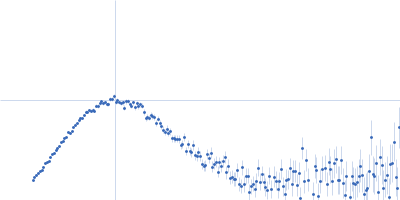
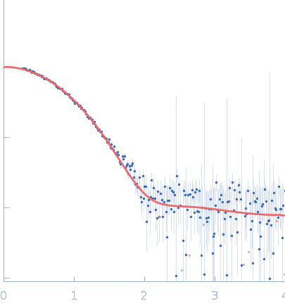
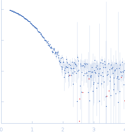
SAXS data from solutions of NanoLuc luciferase in 10 mM Tris-HCl, 50 mM NaCl, pH 7.5 were collected using a Rigaku BioSAXS-2000 at CEITEC - Masaryk University (Brno, Czech Republic) equipped with a Rigaku HyPix-3000 detector at a sample-detector distance of 0.5 m and at a wavelength of λ = 0.154 nm (I(s) vs s, where s = 4πsinθ/λ, and 2θ is the scattering angle). One solute concentration of 3.00 mg/ml was measured at 20°C. One 2400 second frame was collected. The data were normalized to the intensity of the transmitted beam and radially averaged; the scattering of the solvent-blank was subtracted.

Oplophorus-luciferin 2-monooxygenase catalytic subunit (NanoLuc)
Mol. type   Protein
Organism   Oplophorus gracilirostris
Olig. state   Monomer
Mon. MW   20.4 kDa
Sequence   FASTA
 
PDB ID   8AQ6