Structural properties of the HNF-1A transactivation domain

Kind L, Driver M, Raasakka A, Onck P, Njølstad P, Arnesen T, Kursula P, Frontiers in Molecular Biosciences 10 (2023) DOI

SASDSZ8 – The DNA binding domain (DBD) of Hepatocyte nuclear factor 1-alpha (HNF-1A)

Hepatocyte nuclear factor 1-alpha
MWexperimental 14 kDa
MWexpected 23 kDa
VPorod 22 nm3
log I(s) 1.42×10² 1.42×101 1.42×100 1.42×10⁻¹
Hepatocyte nuclear factor 1-alpha small angle scattering data  s, nm-1
ln I(s)
Hepatocyte nuclear factor 1-alpha Guinier plot ln 1.43×10² Rg: 2.7 nm 0 (2.7 nm)-2 s2
(sRg)2I(s)/I(0)
Hepatocyte nuclear factor 1-alpha Kratky plot 1.104 0 3 sRg
p(r)
Hepatocyte nuclear factor 1-alpha pair distance distribution function Rg: 2.8 nm 0 Dmax: 10.2 nm

Data validation


There are no models related to this curve.

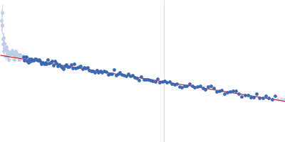
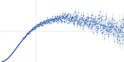
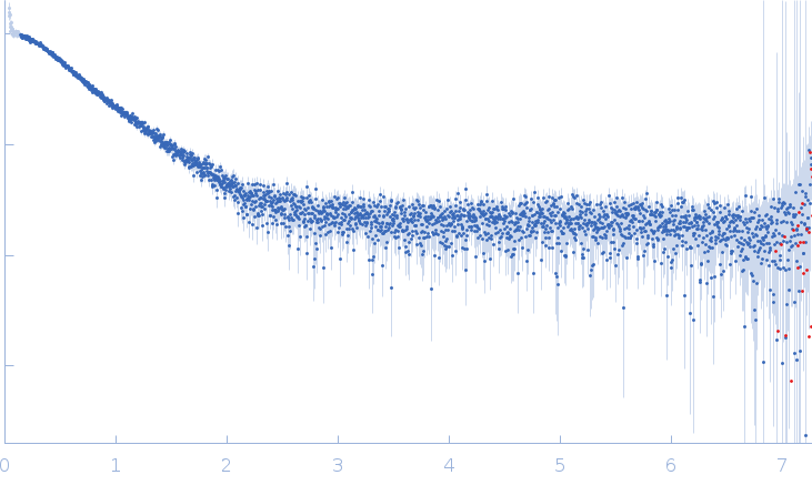
Synchrotron SAXS data from solutions of the DNA binding domain of hepatocyte nuclear factor 1-alpha in 20 mM HEPES, 500 mM NaCl, 1 mM TCEP, pH 8 were collected on the EMBL P12 beam line at PETRA III (DESY; Hamburg, Germany) using a Pilatus 6M detector at a sample-detector distance of 3 m and at a wavelength of λ = 0.124 nm (I(s) vs s, where s = 4πsinθ/λ, and 2θ is the scattering angle). One solute concentration of 2.60 mg/ml was measured at 20°C. 39 successive 0.045 second frames were collected. The data were normalized to the intensity of the transmitted beam and radially averaged; the scattering of the solvent-blank was subtracted.

Bovine serum albumin was used as a standard protein for molecular weight determination. A dilution series (0.5 mg/ml–2.6 mg/ml) was prepared in order to investigate potential concentration-dependent effects.

Hepatocyte nuclear factor 1-alpha (HNF-1A DBD)
Mol. type   Protein
Organism   Homo sapiens
Olig. state   Monomer
Mon. MW   22.8 kDa
 
UniProt   P20823 (83-279)
Sequence   FASTA