Synchrotron WAXS data of poly-histidine tagged Ribose binding protein from Escherichia coli in 50 mM Tris, 50 mM NaCl, 10% glycerol, pH 7 were collected on the BL-15A2 beam line at the Photon Factory (PF), High Energy Accelerator Research Organization (KEK; Tsukuba, Japan) using a Pilatus3 2M detector at a sample-detector distance of 0.5 m and at a wavelength of λ = 0.121297 nm (I(s) vs s, where s = 4πsinθ/λ, and 2θ is the scattering angle). One solute concentration of 16.00 mg/ml was measured at 22°C. 60 successive 10 second frames were collected. The data were normalized to the intensity of the transmitted beam and radially averaged; the scattering of the solvent-blank was subtracted.
Note: The experimental molecular weight, Rg, Dmax and Porod volume are not valid parameters in this case, and are provided only as approximate estimates.
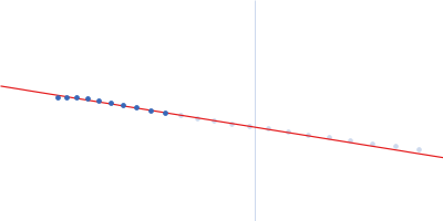
 s, nm-1
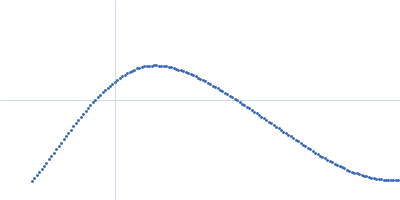