Alternative splicing controls teneurin-3 compact dimer formation for neuronal recognition

Gogou C, Beugelink J, Frias C, Kresik L, Jaroszynska N, Drescher U, Janssen B, Hindges R, Meijer D, Nature Communications 15(1) (2024) DOI

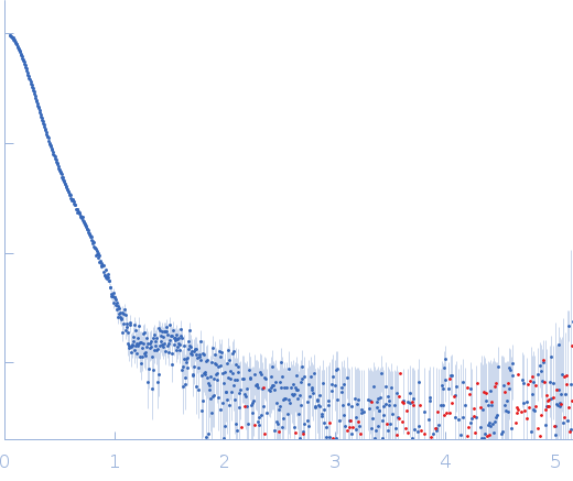
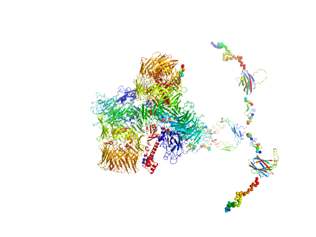
SASDTG3 – Teneurin-3 A1B0 isoform in 2 mM calcium - 0.74 mg/mL

Isoform A1B0 of Teneurin-3
MWI(0) 589 kDa
MWexpected 541 kDa
VPorod 1089 nm3
log I(s) 4.23×10² 4.23×101 4.23×100 4.23×10⁻¹
Isoform A1B0 of Teneurin-3 small angle scattering data  s, nm-1
ln I(s)
Isoform A1B0 of Teneurin-3 Guinier plot ln 4.24×10² Rg: 7.7 nm 0 (7.7 nm)-2 s2
(sRg)2I(s)/I(0)
Isoform A1B0 of Teneurin-3 Kratky plot 1.104 0 3 sRg
Dmax: 30 nm

Data validation


Fits and models


log I(s)
 s, nm-1
Isoform A1B0 of Teneurin-3 CORAL model

Synchrotron SAXS data from solutions of Teneurin-3 A1B0 isoform in 20 mM HEPES, 150 mM NaCl, 2 mM CaCl2, pH 7.8 were collected on the BM29 beam line at the ESRF (Grenoble, France) using a Pilatus3 2M detector at a wavelength of λ = 0.099188 nm (I(s) vs s, where s = 4πsinθ/λ, and 2θ is the scattering angle). One solute concentration of 0.72 mg/ml was measured at 20°C. 10 successive 1 second frames were collected. The data were normalized to the intensity of the transmitted beam and radially averaged; the scattering of the solvent-blank was subtracted.

Sample detector distance = UNKNOWN

Isoform A1B0 of Teneurin-3 (Ten3-A1B0)
Mol. type   Protein
Organism   Mus musculus
Olig. state   Dimer
Mon. MW   270.7 kDa
 
UniProt   Q9WTS6-4 (343-2708)
Sequence   FASTA