|
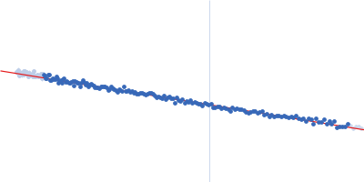
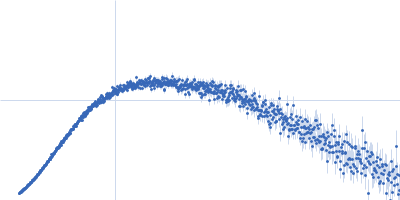
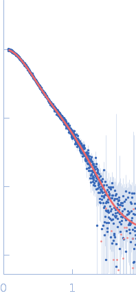
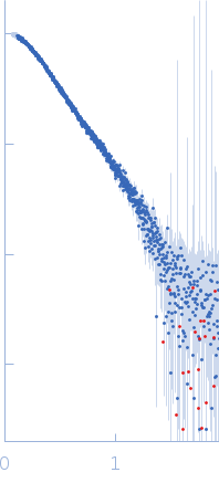
Synchrotron SAXS data from solutions of a GST-TrbB protein fusion in 20 mM HEPES, 100 mM NaCl, 5% glycerol, 0.05% NP40, pH 7 were collected on the BioCAT 18ID beam line at the Advanced Photon Source (APS), Argonne National Laboratory (Lemont, IL, USA) using a Eiger2 XE 9M (Dectris) detector at a sample-detector distance of 3.7 m and at a wavelength of λ = 0.1033 nm (I(s) vs s, where s = 4πsinθ/λ, and 2θ is the scattering angle). In-line size-exclusion chromatography (SEC) SAS was employed. The SEC parameters were as follows: A 300.00 μl sample at 5.5 mg/ml was injected at a 0.60 ml/min flow rate onto a GE Superdex 200 Increase 10/300 column at 20°C. 2594 successive 0.500 second frames were collected. The data were normalized to the intensity of the transmitted beam and radially averaged; the scattering of the solvent-blank was subtracted.
The sample underwent sequential multi-angle laser light scattering analysis using a Wyatt DAWN Heleos II MALS system. NP40 = nonyl phenoxypolyethoxylethanol (https://www.ebi.ac.uk/chebi/searchId.do?chebiId=63016).
|
|
 s, nm-1