Altered protein dynamics and a more reactive catalytic cysteine in a neurodegeneration-associated UCHL1 mutant.

Kenny S, Lai CH, Chiang TS, Brown K, Hewitt CS, Krabill AD, Chang HT, Wang YS, Flaherty DP, Danny Hsu ST, Das C, J Mol Biol :168438 (2024) Europe PMC

SASDTP2 – Ubiquitin carboxy-terminal hydrolase L1 (UCHL1) wild type

Ubiquitin carboxyl-terminal hydrolase isozyme L1
MWexperimental 37 kDa
MWexpected 26 kDa
VPorod 26 nm3
log I(s) 1.28×10⁻¹ 1.28×10⁻² 1.28×10⁻³ 1.28×10⁻⁴
Ubiquitin carboxyl-terminal hydrolase isozyme L1 small angle scattering data  s, nm-1
ln I(s)
Ubiquitin carboxyl-terminal hydrolase isozyme L1 Guinier plot ln 1.29×10⁻¹ Rg: 1.9 nm 0 (1.9 nm)-2 s2
(sRg)2I(s)/I(0)
Ubiquitin carboxyl-terminal hydrolase isozyme L1 Kratky plot 1.104 0 3 sRg
p(r)
Ubiquitin carboxyl-terminal hydrolase isozyme L1 pair distance distribution function Rg: 1.9 nm 0 Dmax: 5.9 nm

Data validation


There are no models related to this curve.

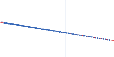
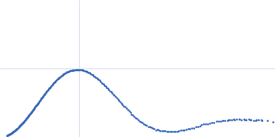
Synchrotron SAXS data from solutions of ubiquitin carboxy-terminal hydrolase L1 in 20 mM HEPES, 150 mM NaCl, 1 mM TCEP, 0.03% NaN3, pH 7.5 were collected on the 13A beam line at the Taiwan Photon Source, NSRRC (Hsinchu, Taiwan) using a Eiger X 1M and Eiger X 9M detector at a sample-detector distance of 1 m and at a wavelength of λ = 0.0827 nm (I(s) vs s, where s = 4πsinθ/λ, and 2θ is the scattering angle). One solute concentration of 14.00 mg/ml was measured at 13°C. Six successive 2 second frames were collected. The data were normalized to the intensity of the transmitted beam and radially averaged; the scattering of the solvent-blank was subtracted.
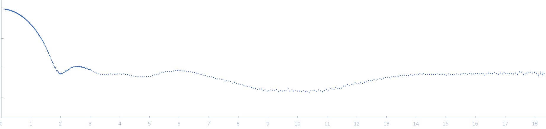
Ubiquitin carboxyl-terminal hydrolase isozyme L1 (UCHL1 WT)
Mol. type   Protein
Organism   Homo sapiens
Olig. state   Monomer
Mon. MW   25.9 kDa
 
UniProt   P09936 (1-223)
Sequence   FASTA