A secreted bacterial protein protects bacteria from cationic antimicrobial peptides by entrapment in phase separated droplets

Ostan N, Cole G Wang F, Reichheld S, Moore G, Pan C, Yu R, Lai C, Sharpe S, Lee H, Schryvers A, Moraes T, PNAS Nexus (2024) DOI

SASDTT7 – Lactoferrin binding protein B

Transferrin-binding protein B
MWexperimental 91 kDa
MWexpected 97 kDa
VPorod 130 nm3
log I(s) 4.35×100 4.35×10⁻¹ 4.35×10⁻² 4.35×10⁻³
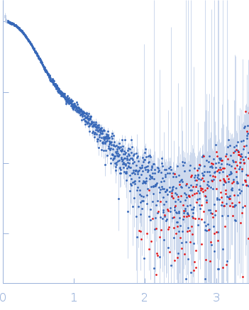
Transferrin-binding protein B small angle scattering data  s, nm-1
ln I(s)
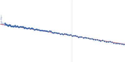
Transferrin-binding protein B Guinier plot ln 4.35×100 Rg: 3.7 nm 0 (3.7 nm)-2 s2
(sRg)2I(s)/I(0)
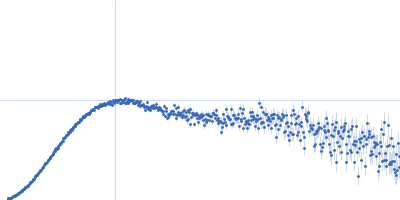
Transferrin-binding protein B Kratky plot 1.104 0 3 sRg
p(r)
Transferrin-binding protein B pair distance distribution function Rg: 3.9 nm 0 Dmax: 12.4 nm

Data validation


There are no models related to this curve.

Synchrotron SAXS data from solutions of lactoferrin binding protein B in 20 mM HEPES, 150 mM NaCl, pH 7.4 were collected on the BioCAT 18ID beam line at the Advanced Photon Source (APS), Argonne National Laboratory (Lemont, IL, USA) using a Pilatus3 X 1M detector at a sample-detector distance of 3.6 m and at a wavelength of λ = 0.1033 nm (I(s) vs s, where s = 4πsinθ/λ, and 2θ is the scattering angle). One solute concentration of 0.60 mg/ml was measured at 22°C. 1800 successive 0.500 second frames were collected. The data were normalized to the intensity of the transmitted beam and radially averaged; the scattering of the solvent-blank was subtracted.

Transferrin-binding protein B (LbpB)
Mol. type   Protein
Organism   Moraxella bovis
Olig. state   Monomer
Mon. MW   96.8 kDa
 
UniProt   K7P8F3 (41-900)
Sequence   FASTA