Inhibition of Mycobacterium tuberculosis UvrB by small molecules: Potent NER disruption and structural insights into dimer conformation.

Jahan F, Anand S, Kumar S, Pandey G, Siddiqi MI, Krishnan MY, Ramachandran R, Int J Biol Macromol :147338 (2025) Europe PMC

SASDTT8 – UvrB (UvrABC system protein B)

UvrABC system protein B
MWexperimental 156 kDa
MWexpected 156 kDa
VPorod 120 nm3
log I(s) 4.54×10³ 4.54×10² 4.54×101 4.54×100
UvrABC system protein B small angle scattering data  s, nm-1
ln I(s)
UvrABC system protein B Guinier plot ln 4.55×10³ Rg: 6.7 nm 0 (6.7 nm)-2 s2
(sRg)2I(s)/I(0)
UvrABC system protein B Kratky plot 1.104 0 3 sRg
p(r)
UvrABC system protein B pair distance distribution function Rg: 5.8 nm 0 Dmax: 17.5 nm

Data validation


Fits and models


log I(s)
 s, nm-1
UvrABC system protein B DAMFILT model

log I(s)
 s, nm-1
UvrABC system protein B HADDOCK model

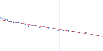
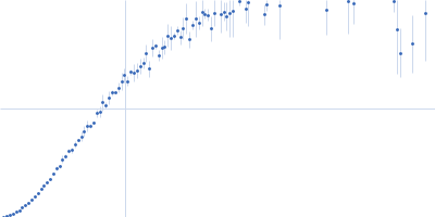
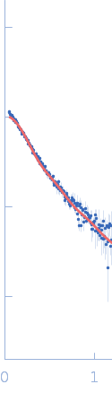
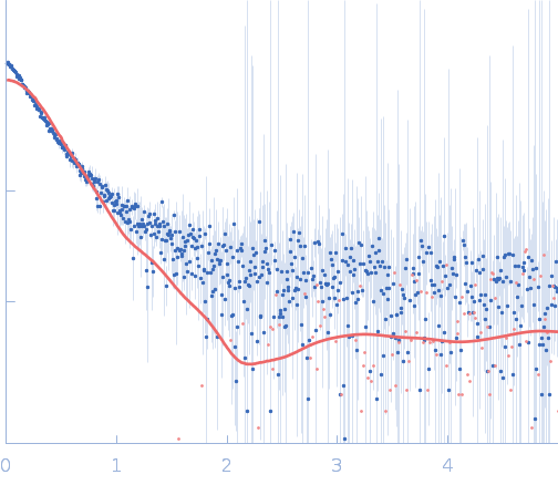
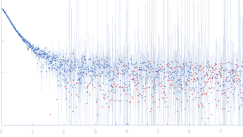
SAXS data from solutions of UvrB (UvrABC system protein B) in 50 mM Tris pH-8, 200 mM NaCl, 2% glycerol were collected using an Anton Paar SAXSpace instrument at the CSIR-Central Drug Research Institute (Lucknow, India) equipped with a Mythen2 R 1K detector at a sample-detector distance of 0.3 m and at a wavelength of λ = 0.154 nm (I(s) vs s, where s = 4πsinθ/λ, and 2θ is the scattering angle). One solute concentration of 5.00 mg/ml was measured at 16°C. Two successive 1800 second frames were collected. The data were normalized to the intensity of the transmitted beam and radially averaged; the scattering of the solvent-blank was subtracted.

CAUTION: Dmax severely underestimated. CAUTION: The DAM model displayed in this entry did not generate the corresponding fit to the SAXS data, but represents the volume and bead-occupancy corrected shape derived from the spatial alignment of an unknown number of individual models.

UvrABC system protein B (UvrB)
Mol. type   Protein
Organism   Mycobacterium tuberculosis (strain ATCC 25618 / H37Rv)
Olig. state   Dimer
Mon. MW   78.0 kDa
 
UniProt   P9WFC7 (22-719)
Sequence   FASTA