Fusion then fission: splitting and reassembly of an artificial fusion-protein nanocage.

Ohara N, Kawakami N, Arai R, Adachi N, Ikeda A, Senda T, Miyamoto K, Chem Commun (Camb) (2024) Europe PMC

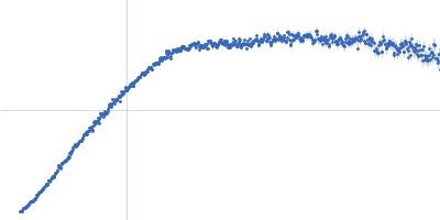
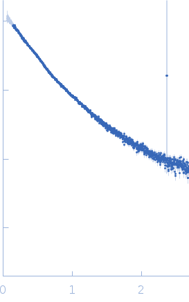
SASDUE2 – TIP120 mutant K26E in CoreC (C-terminal split fragment of a fusion protein that assembles to Truncated Icosahedral protein composed of TIP60 ) subunit with EDTA

N-terminal split fragment of a single subunit of Truncated Icosahedral Protein composed of 60-mer fusion proteins
K26E mutant of C-terminal split fragment of a single subunit of Truncated Icosahedral Protein composed of 60-mer fusion proteins
MWexperimental 48 kDa
MWexpected 1217 kDa
VPorod 110 nm3
log I(s) 1.32×10⁻¹ 1.32×10⁻² 1.32×10⁻³ 1.32×10⁻⁴
N-terminal split fragment of a single subunit of Truncated Icosahedral Protein composed of 60-mer fusion proteins K26E mutant of C-terminal split fragment of a single subunit of Truncated Icosahedral Protein composed of 60-mer fusion proteins small angle scattering data  s, nm-1
ln I(s)
N-terminal split fragment of a single subunit of Truncated Icosahedral Protein composed of 60-mer fusion proteins K26E mutant of C-terminal split fragment of a single subunit of Truncated Icosahedral Protein composed of 60-mer fusion proteins Guinier plot ln 1.33×10⁻¹ Rg: 4.7 nm 0 (4.7 nm)-2 s2
(sRg)2I(s)/I(0)
N-terminal split fragment of a single subunit of Truncated Icosahedral Protein composed of 60-mer fusion proteins K26E mutant of C-terminal split fragment of a single subunit of Truncated Icosahedral Protein composed of 60-mer fusion proteins Kratky plot 1.104 0 3 sRg
p(r)
N-terminal split fragment of a single subunit of Truncated Icosahedral Protein composed of 60-mer fusion proteins K26E mutant of C-terminal split fragment of a single subunit of Truncated Icosahedral Protein composed of 60-mer fusion proteins pair distance distribution function Rg: 4.4 nm 0 Dmax: 13.9 nm

Data validation


There are no models related to this curve.

Synchrotron SAXS data from solutions of TIP120 mutant K26E (CoreC) in 25 mM HEPES, 100 mM NaCl, 4 mM BaCl2, 32 mM EDTA, 5%(v/v) glycerol, pH 8 were collected on the BL-10C beam line at the Photon Factory (PF), High Energy Accelerator Research Organization (KEK; Tsukuba, Japan) using a Pilatus3 2M detector at a sample-detector distance of 2.0 m and at a wavelength of λ = 0.15 nm (I(s) vs s, where s = 4πsinθ/λ, and 2θ is the scattering angle). Solute concentrations ranging between 1.0 and 4.7 mg/ml were measured . 30 successive 5 second frames were collected. The data were normalized to the intensity of the transmitted beam and radially averaged; the scattering of the solvent-blank was subtracted. The low angle data collected at lower concentrations were extrapolated to infinite dilution and merged with the higher concentration data to yield the final composite scattering curve.

Cell temperature = UNKNOWN. Storage temperature = UNKNOWN. Dmax appears underestimated.

N-terminal split fragment of a single subunit of Truncated Icosahedral Protein composed of 60-mer fusion proteins (CoreN)
Mol. type   Protein
Organism   synthetic construct
Olig. state   Other
Mon. MW   6.0 kDa
Sequence   FASTA
 
K26E mutant of C-terminal split fragment of a single subunit of Truncated Icosahedral Protein composed of 60-mer fusion proteins (CoreC K26E)
Mol. type   Protein
Organism   synthetic construct
Olig. state   Other
Mon. MW   14.3 kDa
Sequence   FASTA