Photocobilins integrate B(12) and bilin photochemistry for enzyme control.

Zhang S, Jeffreys LN, Poddar H, Yu Y, Liu C, Patel K, Johannissen LO, Zhu L, Cliff MJ, Yan C, Schirò G Weik M, Sakuma M, Levy CW, Leys D, Heyes DJ, Scrutton NS, Nat Commun 15(1):2740 (2024) Europe PMC

SASDUS3 – Dark-adapted photocobilin diguanylate cyclase fusion from Acidimicrobiaceae bacterium

GGDEF domain-containing protein
MWexperimental 130 kDa
MWexpected 132 kDa
VPorod 194 nm3
log I(s) 1.01×10³ 1.01×10² 1.01×101 1.01×100
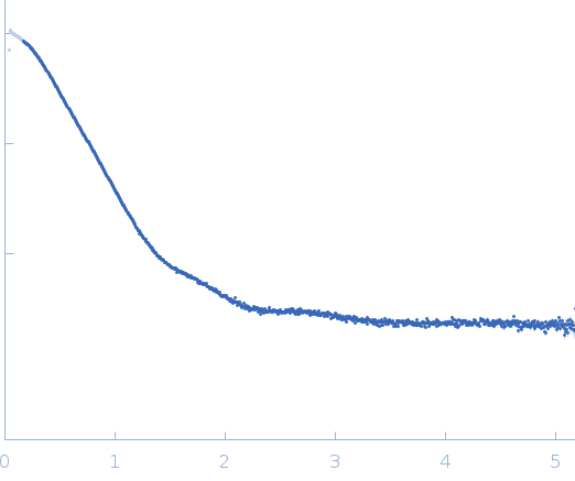
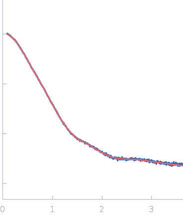
GGDEF domain-containing protein small angle scattering data  s, nm-1
ln I(s)
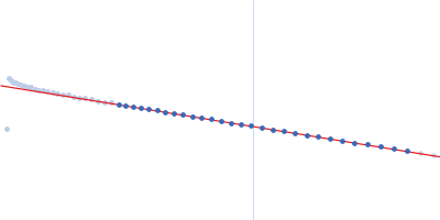
GGDEF domain-containing protein Guinier plot ln 1.02×10³ Rg: 4.1 nm 0 (4.1 nm)-2 s2
(sRg)2I(s)/I(0)
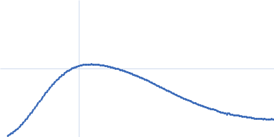
GGDEF domain-containing protein Kratky plot 1.104 0 3 sRg
p(r)
GGDEF domain-containing protein pair distance distribution function Rg: 4.2 nm 0 Dmax: 14.9 nm

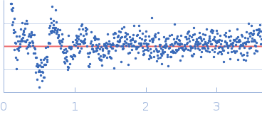
Data validation


Fits and models


log I(s)
 s, nm-1
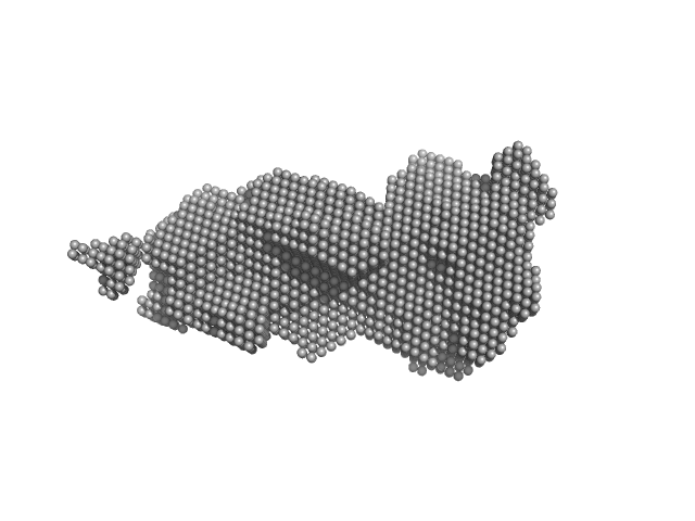
GGDEF domain-containing protein DAMMIF model

Synchrotron SAXS data from solutions of a dark-adapted photocobilin diguanylate cyclase fusion in 20 mM HEPES, 150 mM NaCl, pH 6.8 were collected on the BM29 beam line at the ESRF (Grenoble, France) using a Pilatus3 2M detector at a sample-detector distance of 2.9 m and at a wavelength of λ = 0.099187 nm (I(s) vs s, where s = 4πsinθ/λ, and 2θ is the scattering angle). One solute concentration of 5.00 mg/ml was measured at 20°C. 10 successive 0.990 second frames were collected. The data were normalized to the intensity of the transmitted beam and radially averaged; the scattering of the solvent-blank was subtracted.

GGDEF domain-containing protein
Mol. type   Protein
Organism   Acidimicrobiaceae bacterium
Olig. state   Dimer
Mon. MW   66.1 kDa
 
UniProt   A0A349B205 (1-608)
Sequence   FASTA