Structural flexibility of domains in human AOC3

Gabriela Guedez.

SASDUU3 – Human primary amine oxidase copper-containing 3 (AOC3)

Membrane primary amine oxidase
MWexperimental 224 kDa
MWexpected 164 kDa
VPorod 372 nm3
log I(s) 3.56×10⁻² 3.56×10⁻³ 3.56×10⁻⁴ 3.56×10⁻⁵
Membrane primary amine oxidase small angle scattering data  s, nm-1
ln I(s)
Membrane primary amine oxidase Guinier plot ln 3.57×10⁻² Rg: 4.6 nm 0 (4.6 nm)-2 s2
(sRg)2I(s)/I(0)
Membrane primary amine oxidase Kratky plot 1.104 0 3 sRg
p(r)
Membrane primary amine oxidase pair distance distribution function Rg: 4.2 nm 0 Dmax: 11.7 nm

Data validation


Fits and models


log I(s)
 s, nm-1
Membrane primary amine oxidase DAMMIN model

log I(s)
 s, nm-1
Membrane primary amine oxidase PYMOL model

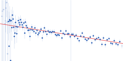
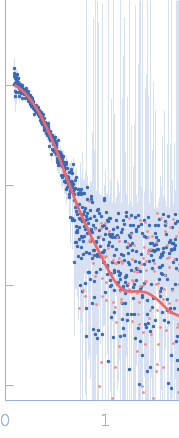
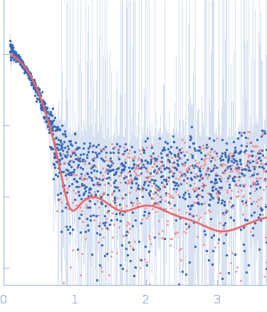
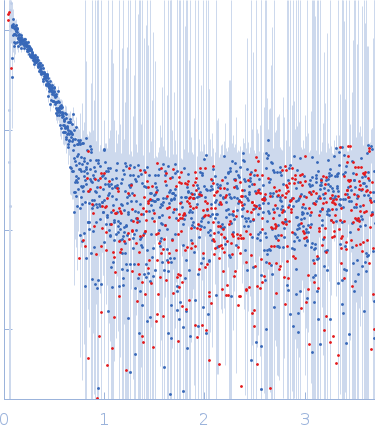
Synchrotron SAXS data from solutions of human AOC3 dimer in 137 mM NaCl, 2.7 mM KCl, 10 mM Na2HPO4, 1.8 mM KH2PO4, pH 7.4 were collected on the B21 beam line at the Diamond Light Source (Didcot, UK) using a Pilatus 2M detector at a sample-detector distance of 4 m and at a wavelength of λ = 0.1 nm (I(s) vs s, where s = 4πsinθ/λ, and 2θ is the scattering angle). Solute concentrations ranging between 0.3 and 0.5 mg/ml were measured at 25°C. 27 successive 10 second frames were collected. The data were normalized to the intensity of the transmitted beam and radially averaged; the scattering of the solvent-blank was subtracted and analysed using Primus to produce the data displayed in this entry.

The entry shows 1) the individual ab initio reconstruction and 2) fitting of the experimental scattering curve to the AOC3 crystal structure (PDB entry 8S1Z).

Membrane primary amine oxidase (AOC3)
Mol. type   Protein
Organism   Homo sapiens
Olig. state   Dimer
Mon. MW   81.8 kDa
 
UniProt   Q16853 (27-763)
Sequence   FASTA