|
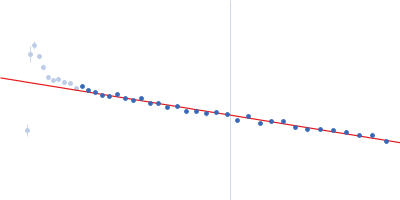
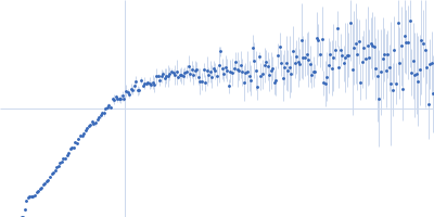
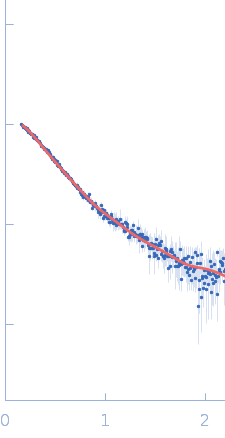
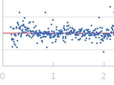
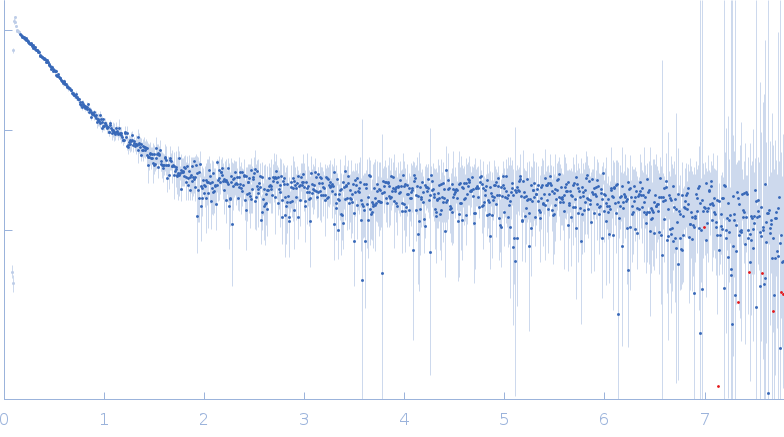
SAXS data from solutions of DNA ligase-1 in 50 mM HEPES pH 7.5, 200 mM NaCl were collected using an Anton Pair SAXSpace instrument at the CSIR-Central Drug Research Institute (Lucknow, India) equipped with a Mythen2 R 1K detector at a sample-detector distance of 0.3 m and at a wavelength of λ = 0.154 nm (I(s) vs s, where s = 4πsinθ/λ, and 2θ is the scattering angle). One solute concentration of 2.00 mg/ml was measured at 10°C. Two successive 30 second frames were collected. The data were normalized to the intensity of the transmitted beam and radially averaged; the scattering of the solvent-blank was subtracted.
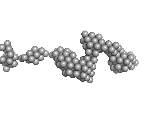
CAUTION!! The Porod volume (~53 nm³) and molecular weight estimates calculated from the SAXS data (22-43 kDa) in addition to the volume (~56 nm³) and molecular weight of the DAM model (~28 kDa) are significantly lower than expected for a protein of 100 kDa.
|
|
DNA ligase 1
|
| Mol. type |
|
Protein |
| Organism |
|
Homo sapiens |
| Olig. state |
|
Monomer |
| Mon. MW |
|
101.7 kDa |
| Sequence |
|
FASTA |
| |
|

 s, nm-1